International Journal of Marine Science, 2017, Vol.7, No.24, 229-246
232
bottom. There was rapid and mass death of fish. Also, grossly focal erosion areas were seen on the operculum and
caudal tail. Deformity in vertebral column. The affected skin showed friable skin (velvet like appearance),
darkened, excessive mucous secretions. Also, sometimes grossly focal erosion caudal tail. Internally the infested
fish, showed pale liver enlarged spleen and fatty position on gastro intestinal tract.
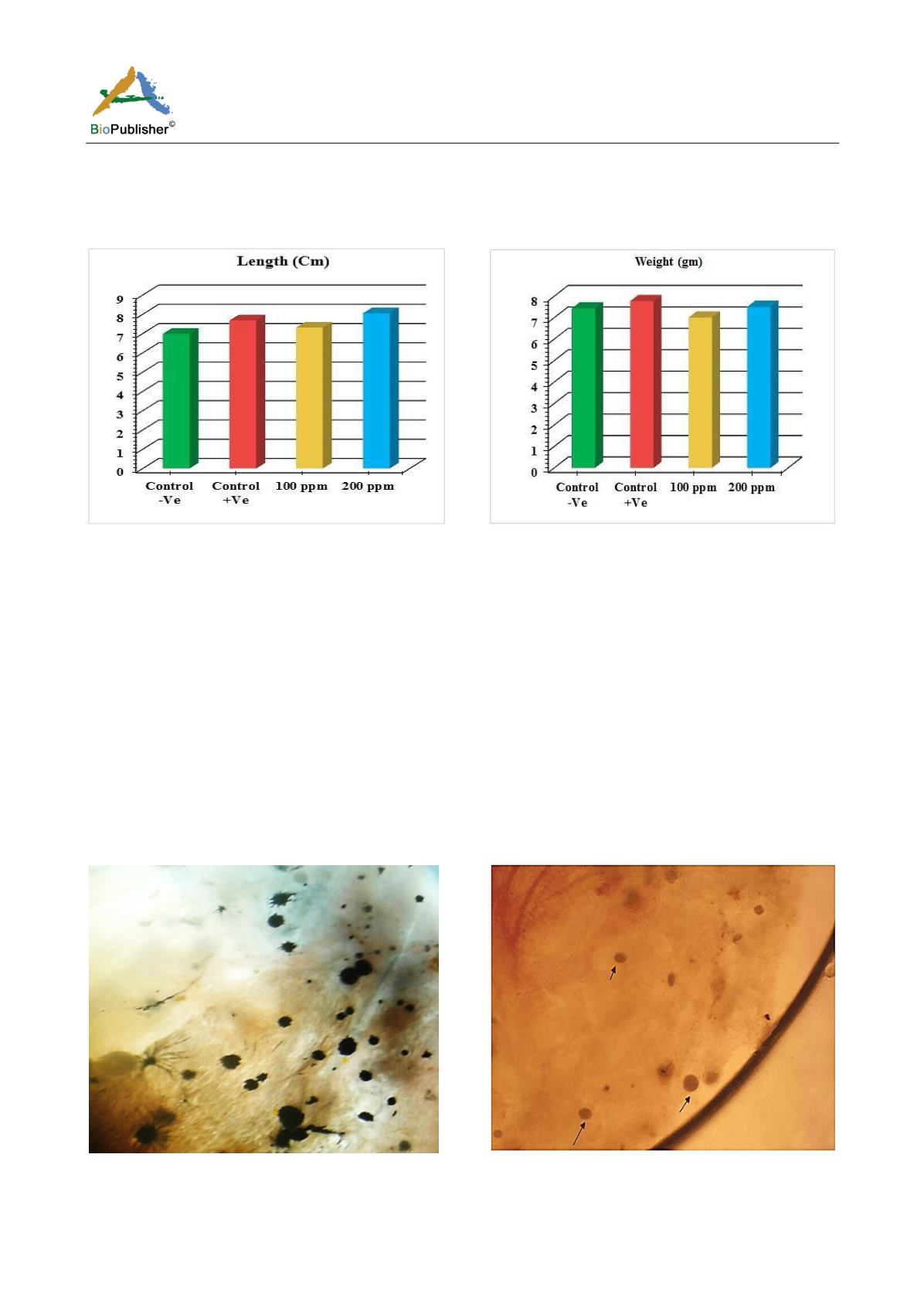
Figure 1 Comparison of Length among treated groups No. (%)
after (30 min.) treatment
Figure 2 Comparison of Weight (gm) among treated groups
after (30 min.) treatment
2.2 Parasitological examination
2.2.1 Morphological description
Microscopic examination of skin and gills scrapings of examined
D. labrax
showed, round to oval small dark
brown mucoid
Amyoodinium ocellatum
stage measured up to 150 μm. Different developmental life stages of
Amyloodinium ocellatum
were seen in the gill and skin tissues as the two main inhabitant organs. The first stage
was trophonts with its root - like structure (Rhizoids). They showed distended appearance with the presence of
feeding stage trophonts dark brown color lodged between the gill filaments. Also, observed between the skin and
fin surface the rhizoids root-like structure (that penetrates deep in to epithelium causing substantial damage to
tissue at the attachment site) of the attached organelle was sometimes visible
as
in figures (Figure 3; Figure 7;
Figure 8; Figure 10)
.
The second was tomont stage; trophont stage feeds for several days, detaches, retracts its
rhizoids and becomes tomont. Tomonts (reproductive stage) in division were occasionally observed as
in
(Figure 3;
Figure 4; Figure 5; Figure 6; Figure 9; Figure 10; Figure 11) and a special picture of advanced stage of division of
tomont stage in Figure 12.
Figure 3 Showing fresh mount of gill tissue heavy infested
with Amyloodinium ocellatum ( trophont stage with rhizoid
root) and Tomont stage( first division.)
Figure 4 Showing fresh mount of gill tissue slightly infested
with Amyloodinium ocellatum ( trophont stage).division


