International Journal of Marine Science, 2017, Vol.7, No.24, 229-246
239
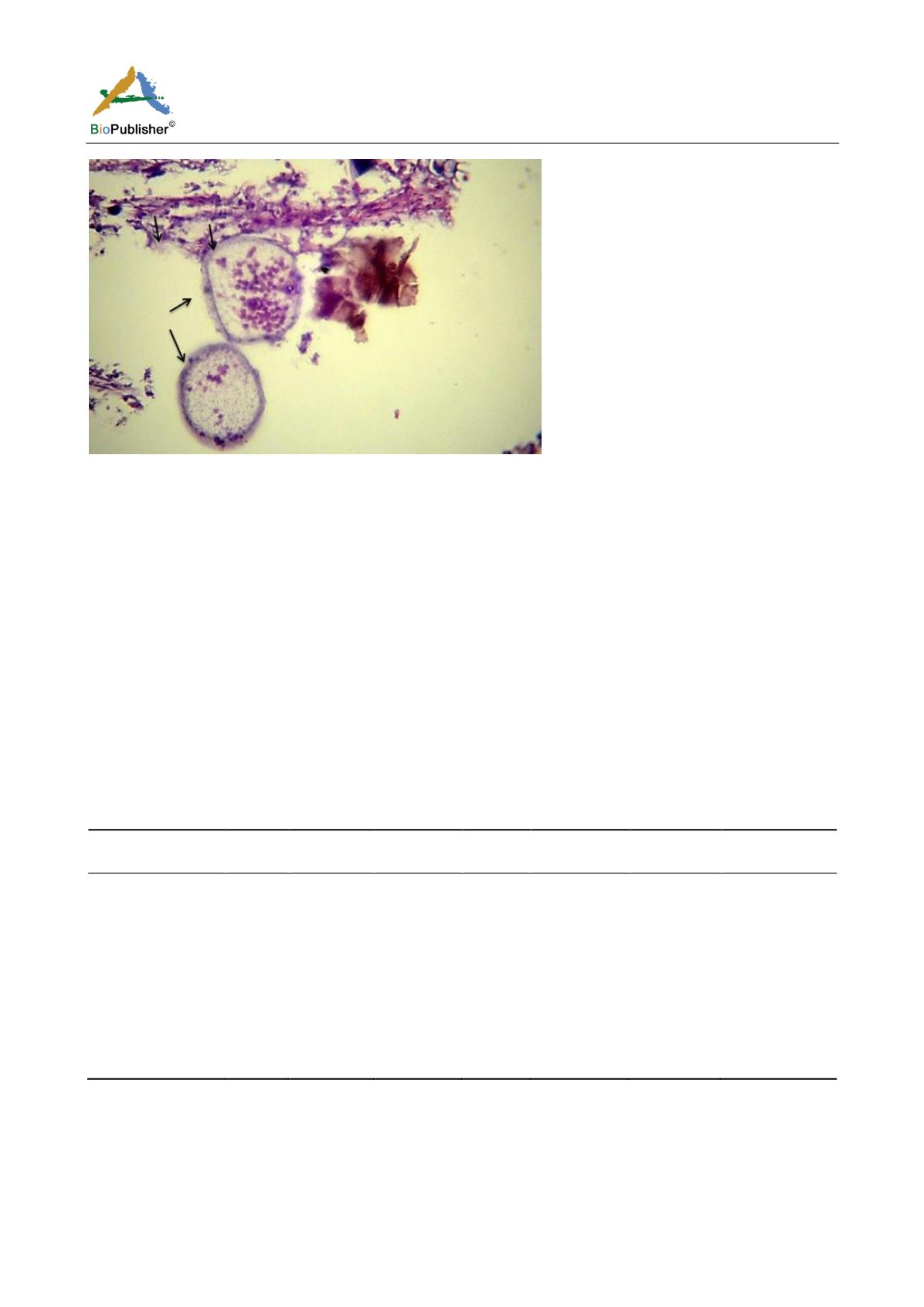
Figure 27 Showing Amyloodinium ocellatum trophont (Arrowheads) and its attachment site to the destructed secondary gill lamella
(Arrow) H&E X400
2.5 Treatment trials for
Amyloodiniosis
Results
2.5.1 Clinical signs before and after treatment
Before treatment the infected fish showed signs of distress loss of appetite flashing behavior mass mortality
accumulation at water surface. Microscopic examination of skin and gill filament showed presence of tomont
stage dark-brown in color mechanically dislodged between gill filaments.
After treatment feed consumption returned to a normal rate fishes in two treated groups resumed feeding from
second day of commencement of treatment. Fish stopped dying increase vitality and trophont count decreased
significally following treatment.
Effects of treatments on detachment of the trophonts and recovery of fish were studied the detachment of
trophonts and its numbers were assessed by examining the gill filament skin and fin swab from the fish after
treatment as in Table 5. The final mean of mortality percentage in each treated group was calculated on
termination of treatment as in Table 4.
Table 4 Showing the final mean of mortality percentage in each treated group that was calculated on termination of treatment
Parameter Group
Total No
Mean Length
(cm)
Mean Weight
(gm)
Dose
Duration of
Exposure
Mean No of
Mortality
Mean % Mortality
First Week
G1 control (-)
30
6.98
7.43
-
-
0
0%
G2 control (+)
30
7.68
7.77
-
-
1
3.33%
G3 Treated
30
7.31
5.13
100 PPm 30 min
1
3.33%
G4 Treated
30
8.05
6.33
200 PPm 30 min
0
0%
Second Week
G1 control (-)
30
6.98
7.43
-
-
0
0%
G2 control (+)
30
7.68
7.77
-
-
3
10%
G3 Treated
30
7.31
5.13
100 PPm 30 min
0
0%
G4 Treated
30
8.05
6.33
200 PPm 30 min
0
0%
Note: Percents within the same column sharing the same subscript are not significantly different at p< 0.05 in the two-sided test
Z-test. Tests are adjusted for all pairwise comparisons using the Bonferroni correction
As shown in Table 4 and Table 5, the mortality rate was decreased in the treated groups G1 and G2 to become
none in the second week. Also, the intensity of infection was decreased in the treated groups where skin smear and
gill smear shown decreased no of
Amyloodinium
than the non-treated group. Therefore, the prevalence rate was
lower in the treated groups than non-treated. It was cleared that, this treatment with was more effective and


