International Journal of Marine Science, 2017, Vol.7, No.24, 229-246
242
fish that were to be treated with 200 ppm showed a mean of 10 ±0.93 trophonts per gill biopsy in first week & 3 ±
1.03 trophonts per skin smear. The fish were retreated with 200 ppm hydrogen peroxide for thirty minute for
another 6 days; the count was down to 4 ±0.93 trophonts per gill biopsy & 2 ±0.97 trophonts per skin smear.
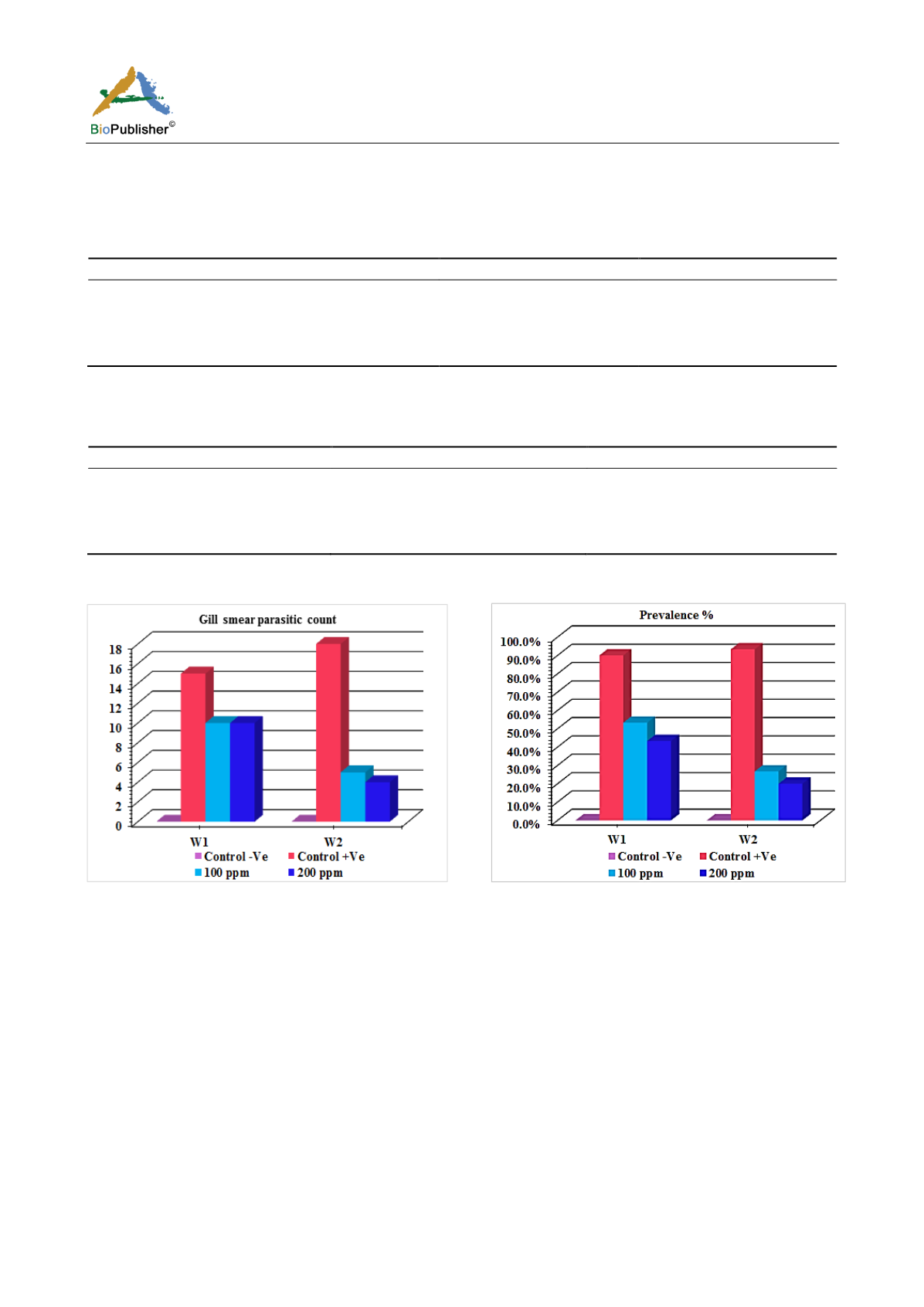
Table 8 Comparison of gill smear parasitic count among treated groups after (30 min.) treatment (G: group; W: week)
W1
W2
Control –Ve(G1)
-
-
Control +Ve(G2)
15±0.74
a
18±0.93
a
100 ppm (G3)
10±1.01
b
5±0.93
b
200 ppm(G4)
10±0.93
b
4±0.93
b
Note: Means within the same column carrying different superscripts are sig. different at P < 0.05 based on Tukey's Honestly
Significant Difference (Tukey’s HSD)
Table 9 Comparison of prevalence % among treated groups after (30 min.) treatment (G: group; W: week)
W1
W2
Control –Ve(G1)
0 (0.0%)
0 (0.0%)
Control +Ve(G2)
27 (90.0%)
a
28 (93.3%)
a
100 ppm (G3)
16 (53.3%)
b
8 (26.7%)
b
200 ppm (G4)
13 (43.3%)
b
6 (20.0%)
b
Note: Percents within the same column not sharing the same subscript are significantly different at p< 0.05 in the two-sided test
Z-test. Tests are adjusted for all pairwise comparisons using the Bonferroni correction
Figure 30 Comparison of gill smear parasitic count among
treated groups after (30 min.) treatment
Figure 31 Comparison of prevalence % among treated groups
after (30 min.) treatment
3 Discussion
Eissa (2002) reported that, by increasing intensification of fish production and lack of health management
measures have led to many disease problems. About 80% of fish disease is parasitic especially in warm water fish.
The ectoparasitic protozoal diseases (gill and skin parasites) of fishes play an effective role in the economic losses
of fish farms through mortality and/or decrease growth rate of fish especially in the highly intensified systems.
Pereira et al. (2010) reported that among the most important ectoparasitic protozoa is
Amyloodinium ocellatum
, a
dinoflagellate which causes one of the most serious diseases of warmwater marine aquaculture and the disease
caused by this organism is commonly referred to as
amyloodiniosis
or marine velvet disease. Lom and Dikov
(1992) reported changes in fish behavior, with jerky movements, swimming at the water surface and decreased
appetite. Levy et al. (2007) suggested that, the first indication of an
amyloodinium
infection is dead or dying fish.
Behavioral signs may include a decrease in or complete lack of feeding activity, flashing (rubbing against objects
in the tank or on the bottom substrate) and coughing (back flushing water across the gills). The skin of heavily
infected fish may have a dull gold or brown sheen. And also may reveal scale loss and patchy accumulation of


