International Journal of Marine Science, 2017, Vol.7, No.24, 229-246
235
Table 2 Showed Prevalence and seasonal dynamics of infestation by
Amyloodinium ocellatum
of European Seabass
Dicentrarchus
labrax
. in 2015-2016
Season
No. of Fish Examined No. of Fish Infested
Prevalence %
species
Infected organs
Spring
546
492
90.10%
Fry
G.S
Summer
222
75
33.78%
Fry
G.S
Autumn
219
108
49.32%
fingerling
G.S
Winter
78
24
30.77%
Adults
G
Total
1065
618
58.02%
2
Calc.
73.55
P-value
< 0.0001
Note: G: gills; S: Skin
Table 3 Correlation among water quality parameters and parasitic infestation during prevalence period of
Amyloodinium ocellatum
infection
Parameter
r
P-value
Temperature
-0.165
0.488
Dissolved Oxygen
0.009
0.97
pH
-.457*
0.043
Salinity (ppt)
.473*
0.035
Ammonia (ppm)
0.057
0.81
Note:
Correlation is significant at the 0.05 level (2-tailed). Only the pH and salinity were strong correlated with A. ocellatum
infestation. Between the parasitic infestation and pH there was an inverse relationship that the result of Pearson Correlation
coefficient test was (-.457) carrying a negative sign. While between the parasitic infestation and salinity there was appositive
relationship that the result of Pearson Correlation coefficient test was (.473) carrying a positive sign
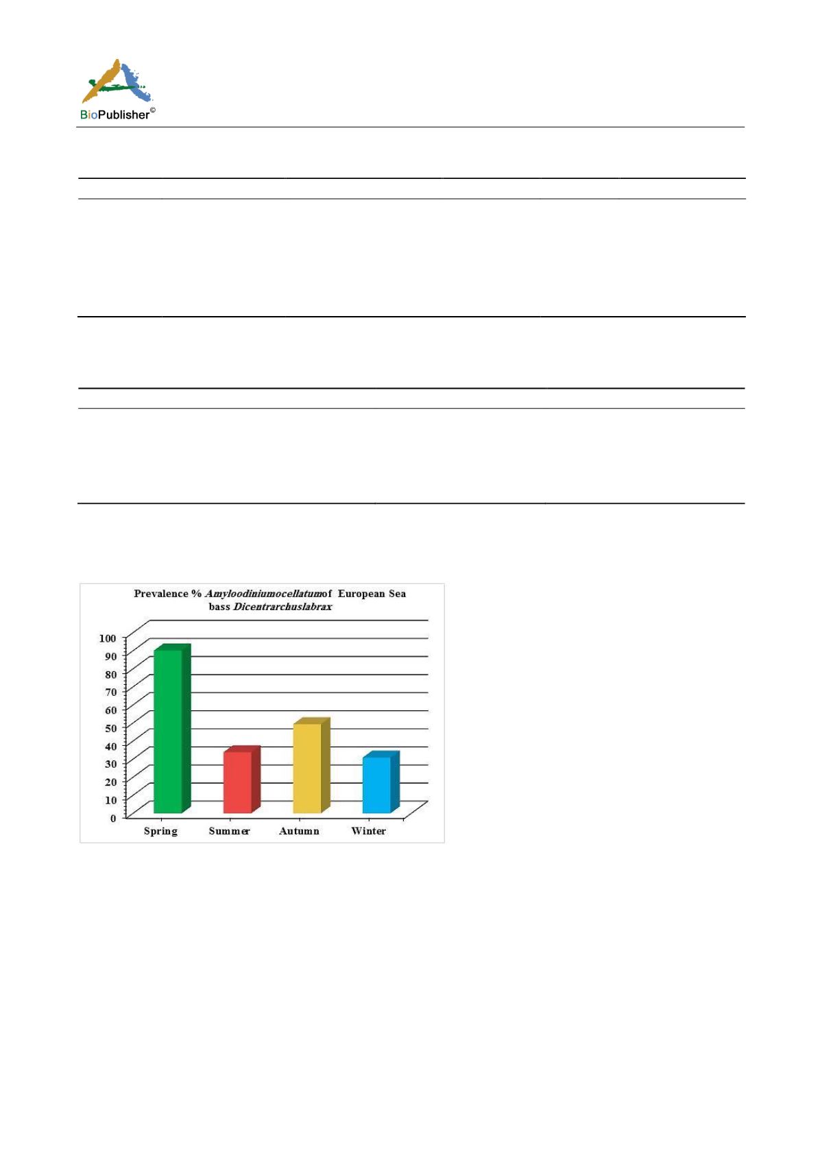
Figure 13 Prevalence and seasonal dynamics of infestation by Amyloodinium ocellatum of
Dicentrarchus labrax.
2.4 Histopathological alterations
Infestations of
D. labrax by Amyloodinium ocellatum
are usually involve the gill as the primary site of infestation
may also the skin and eyes. Thus there were histopathological changes ranged between mild infestations by low
number of trophonts per gill filament and cause little alteration. But, heavy infestations by high number of
trophonts can cause serious gill hyperplasia, inflammation, hemorrhage, and necrosis. As shown in (Figures 19;
Figure 20; Figure 25). Trophonts were also seen inside gill arch and tissues. Concerning the mild pathological
changes,
Amyloodinium ocellatum
trophont was attached to the skin and causing moderate hyperplasia and
desquamation of the covering epithelium (Figure 23; Figure 26). On the other hand, several histpathological
changes were seen in several forms as follow:
Amyloodinium ocellatum
trophonts causing severe hyperplasia in the
gill epithelia and fusion of the secondary gill lamell Figures (Figure 21; Figure 22).
Amyloodinium ocellatum
trophont and its attachment site to the destructed secondary gill lamella (Figure 24; Figure 27).


