Animal Molecular Breeding 2016, Vol.6, No.4, 1-10
6
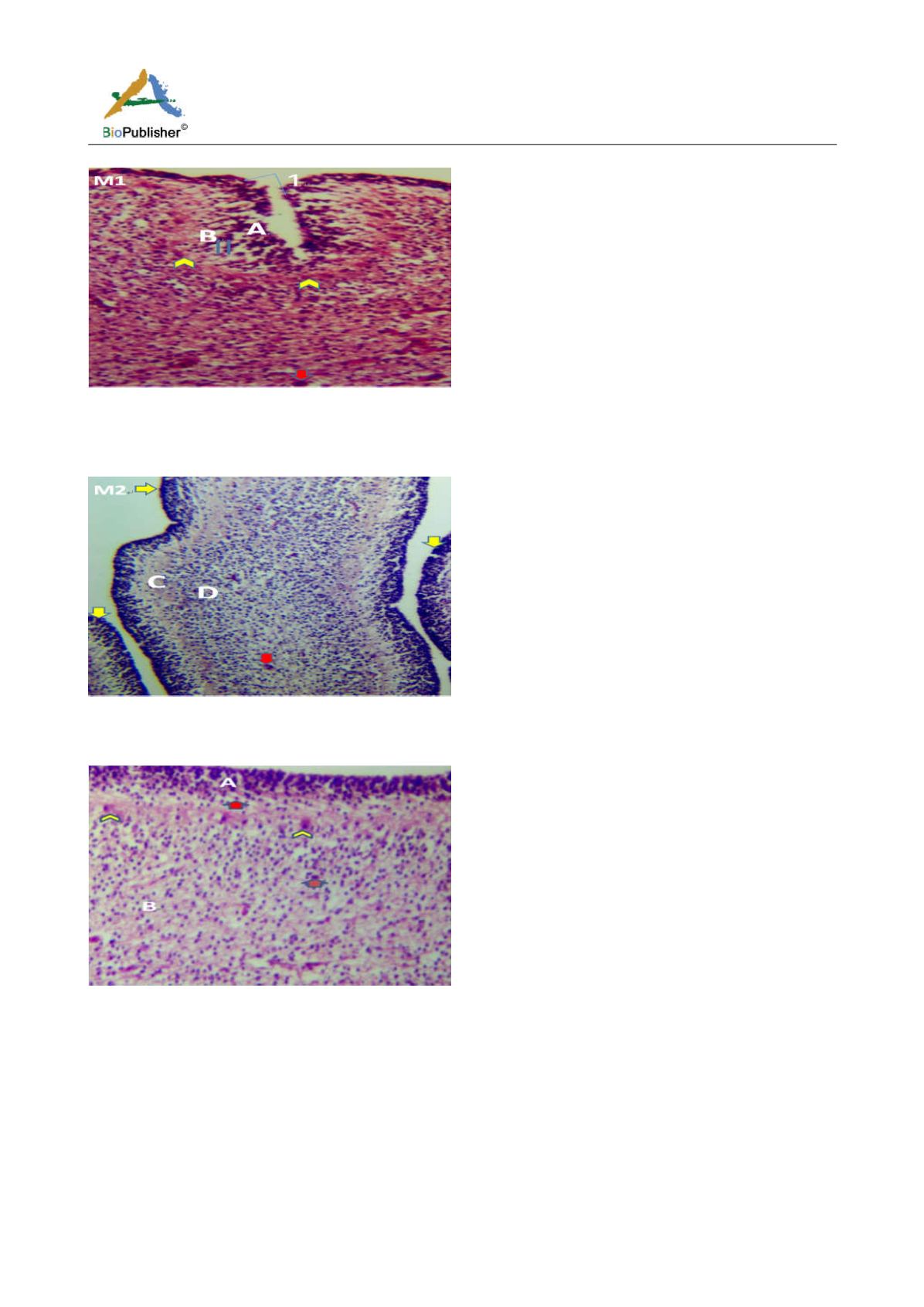
Figure 6a Cerebellum showing, A; External granular layer, B; Primitive molecular layer, 1; Folia deepen, Double arrow; Granule cells
migrating from EGL, Red arrow; Large migratory cells, Arrowheads; Future Purkinje cells, Yellow arrow; Folia formed. (Day 18
pre-hatch). H&E. Magnification X140
Figure 6b Cerebellum showing, A; External granular layer, C; Inner cortical layer, Yellow arrows; Folia formation, Red arrows; Large
migratory cells, Dinner granular layer, (day 20 pre-hatch). H&E. Magnification, X140
Figure 7 Cerebellum showing, A; External granular layer, B; Dark stained inner granular cells, Red arrow; granule cells migrating from EGL, Arrow
heads; Future Purkinje cells, Ox blood arrows; Large granular cells of inner cortical layer. (Day 20 of incubation). H&E. Magnification X140
The cerebellum at day 23 was observed to have numerous large immature migratory cells emerging from the dark
stained granule cells toward the inner cortical layer. The molecular layer began to appear, numerous neuropils and
future Purkinje cells became attached to the neurofibres (Figures 8 and 9). The molecular, granular and Purkinje
layers were distinct at day 27 pre-hatch with the presence of immature Purkinje cells arranged in a row and traces
of external granular layer still prominent.


