Animal Molecular Breeding 2016, Vol.6, No.4, 1-10
7
Figure 8 Cerebellum showing, A; Dense dark stained cell of EGL, B; Molecular layer, C; Granular cell layer, D; White matter, Arrowheads; Larger
migratory cells, Arrows; Migratory Purkinje cells, Arrows; Immature Purkinje cells, (Day 23 of incubation). H&E. Magnification X140
Figure 9 Cerebellum HGF showing, A; EGL, B; Molecular layer, C; Granular layer, Arrowhaeds; Neuropils, Arrows; Migratory Purkinje cells. (Day
24 of incubation). H&E. Magnification X140
Cellular organization at the fourth week post-hatch showed that the large perikarya Purkinje cells were round,
pear-shaped an arranged in single row at the junction between the molecular and granular layers. The cells
demonstrated prominent nucleoli and nuclear materials. Different sizes and shapes of various neurones were
observed at 8th week. The inner granular layer was observed to contain numerous small round granule cells giving
the appearance of tightly packed chromatic nuclei. The white matter was seen to have sparse granule cells with
dense nerve fibres (Figures 10a and 10b) and (Figure 11).
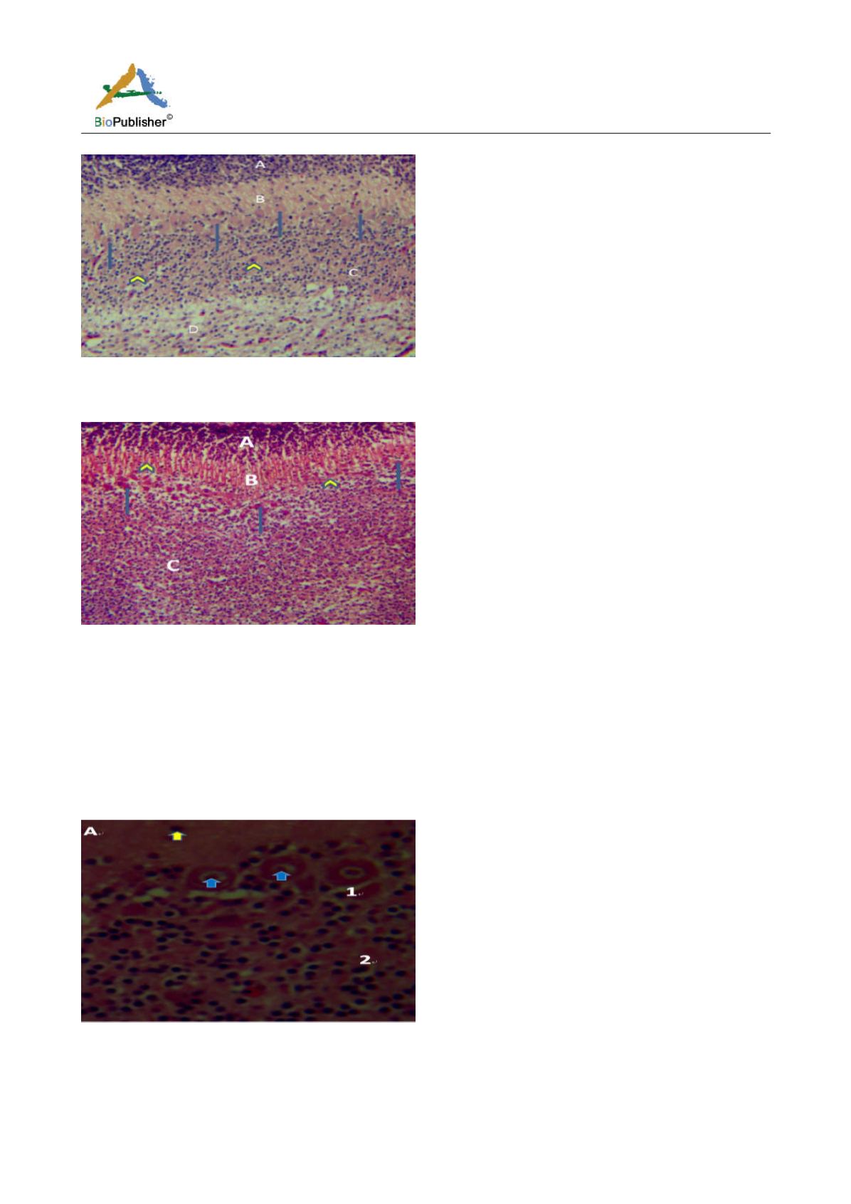
Figure 10a Cerebellum of the GF showing, 1; Purkinje cell, 2; Fibre tracts, Yellow arrow; Basket cell, Blue arrow; Nuclei of Purkinje
cell, (Week 4 Post-hatch). H&E, Magnification; X400


