Animal Molecular Breeding 2016, Vol.6, No.4, 1-10
4
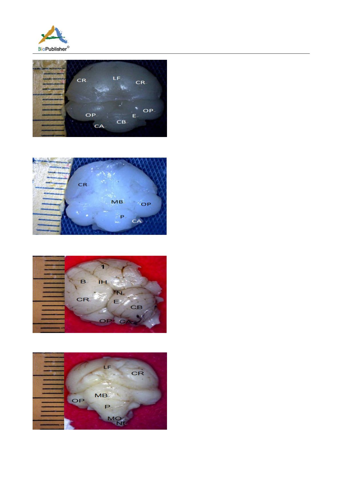
Figure 2a Brain showing cerebellum, CR; Cerebrum, LF; Longitudinal fissure, E; Transverse fissures, OP;
Optic lobe, CB; Cerebellum, CA; Cerebellar auricle (Day 18 of incubation). Magnification X1000
Figure 2b Brain ventral view showing, CR; Cerebrum, MB; midbrain, OP; Optic lobe, P; Pons, CA; Cerebellar auricle (Day 18 of
incubation). Magnification X1000
Figure 3a Whole brain indicating; CR; Cerebrum, IH; Interhemispheric fissure, N; Notch, E; Transverse fissures, OP; Optic lobe, CB;
Cerebellum, CA; Cerebellar auricle, SP; Spinal cord, (Day 27 of incubation). Magnification X1000
Figure 3b Whole brain indicating; CR; Cerebrum, LF; Longit dinal fissure, MB; Midbrain, OP; Optic lobe, P; Pons, Medulla
oblongata, NF; Nuchal flexure. (Day 27 of incubation) Magnification X1000


