Animal Molecular Breeding 2016, Vol.6, No.4, 1-10
3
slipped using diphynylpthalate propylene xylene (DPX) as mountant (Drury and Willington, 1980).
Sections were stained with hematoxylin and eosin (H&E), and cresyl fast violet (CFV) for nuclei and
photomicrographs of sections were taken using digital eyepiece (Scopetek DCM 500, Resolution: 15 Pixels, made
in China) attached to a light microscope (OLYMPUS XSZ107BN, Hamburg, Germany).
3 Results
3.1 Gross features
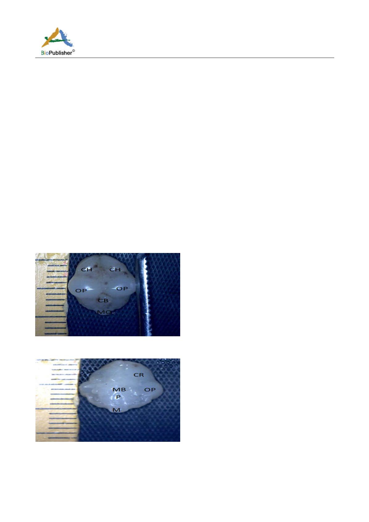
The result from this study revealed that during the pre-hatch period, the cerebellum of the grey breasted helmeted
guinea fowl (HGF) was observed to develop on the dorsal surface of the pons by the 11th day with the cerebrum,
optic lobe, midbrain and medulla oblongata being formed just at the caudal end of the longitudinal fissure,
(Figures 1a and 1b). By day 18 of incubation, there was an increased in the size, shape and location of the
cerebellum. On this same day, fissures of the vermis first appeared showing various lobes which progressively
became distinct. The cerebellar auricles was observed to be attached to the cerebellum at the ventromedial aspects.
The rostral part of the cerebellum tapered to fits into the notch that form part of the transverse process (Figures 2a
and 2b). The size of the cerebellum tends to increase with increase in size and shape of the cerebral hemispheres at
day 27 pre-hatch. Foliation became prominent, rostral part of the cerebellum was broaden and was opposite to
what was seen at day 18 pre-hatch. The rostral part became firmly attached to the cerebral hemispheres and optic
lobes. Dorsally, the middle cerebellar portion was relatively larger, somewhat spherical to convex but tapered
caudally as it joined the pons ventrally. Cerebellar auricles became prominent when viewed from the dorsal and
ventral aspects of the brain. The cerebral hemispheres were large and lissencephalic throughout pre and post-hatch
but bore some surface structures, such as grooves and eminences, at day 27 (Figures 3a and 3b).
Figure 1a Brain dorsal view showing cerebellum, CH; Cerebral hemisphere, OP; Optic lobe, CB; Cerebellum, M; Medulla oblongata,
Magnification, X1000. (Day 11 pre-hatch)
Figure 1b Brain showing ventral view, CR; Cerebrum, MB; Midbrain, OP; Optic lobe, P; Pons, M; Medulla oblongata. Magnification,
x1000, (Day 11 pre-hatch)


