Animal Molecular Breeding 2016, Vol.6, No.4, 1-10
5
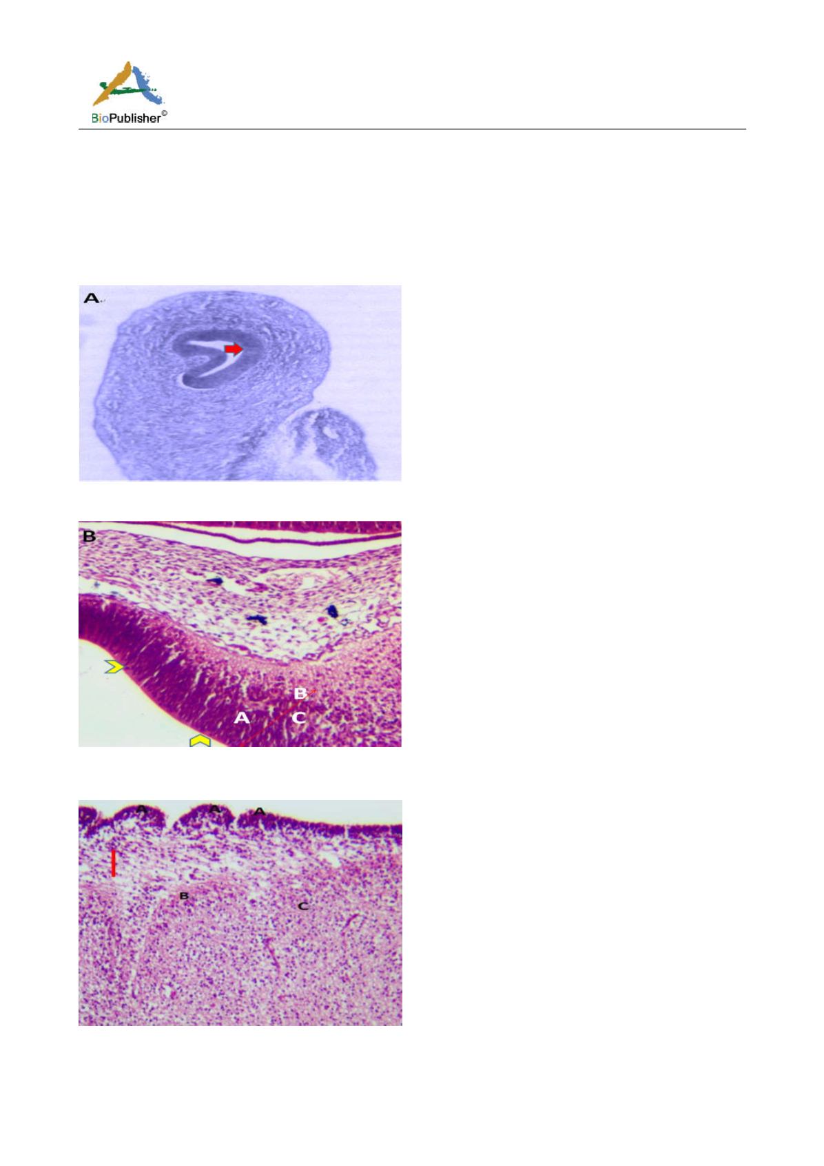
3.2 Histological features
Microanatomical observations on day 11 pre-hatch suggested that the primordium of the cerebellum had been
formed and was made up of cerebellar anlage composed of inner and outer mantle layers. Cells within these regions
or layers migrated from the ventricular neuroepithelium of the primordium (Figures 4a and 4b). Foliation was
observed for the first time on day 13 pre-hatch with granule cells migrating from the external granular layer. The
marginal layer was also interposed with migratory cells, (Figure 5). Foliation was completed at this stage and future
Purkinje cells were also aligned at the inner cortical layer (Figures 6a and 6b) and (Figure 7).
Figure 4a Cerebellum Primordium showing, Arrow: Cerebellar primordium. (Day 11 pre-hatch), Magnification X 140
Figure 4b Cerebellar Primordium showing, A; Inner B; Outer mantle, C; Cerebellar anlage, Arrowheads; ventricular neuroepithelium,
(Day 11 pre-hatch)
Figure 5 Cerebellum showing, A; Folia formation, B; Marginal layer, C; Inner cortical layer, Arrow; Granule cells migrating from
EGL, Day 13 of incubation. H&E. Magnification X140


