International Journal of Clinical Case Reports 2018, Vol.8, No.2, 5-9
6
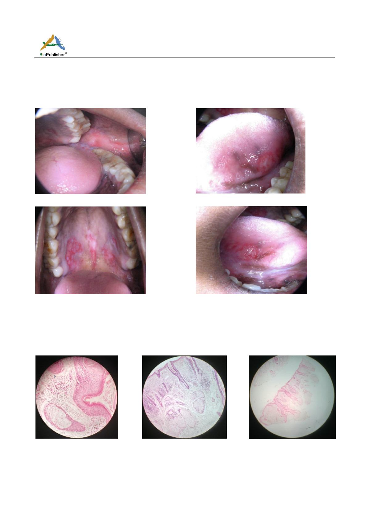
Intraoral examination revealed reddish ulcers on the buccal mucosa, tongue, soft and hard palate (Figure 2, Figure
3, Figure 4 and Figure 5). Patient had visited a private dentist for the same and was prescribed mouthwash and
anti-inflammatory drugs with vitamins supplements but the lesion was not responding to any drug. Our
differential diagnosis was Lupus Erythematosus and Lichen Planus. Patient was advised investigations like routine
hemogram, Xray of the bone, and biopsy with anti-nuclear antibody (A.N.A) testing immuno-fluoroscence.
Figure 2 Lesion on the left buccal mucosa
Figure 3 Ulcer on the ventral surface of tongue
Figure 4 Ulcers on the hard and soft palate
Figure 5 Shallow ulcer on Ventral surface of the tongue
A biopsy of the representative lesion was taken under Local anesthesia under antibiotic coverage. Hemotoxylin
and Eosin (H.E) stain section showed stratified squamous epithelium with hyperkeratosis and acanthosis (Figure
6), perivascular cuffing, liquefaction degeneration of basal cell layer (Figure 7) and chronic inflammatory cells as
well as sub-epithelial vesicles (Figure 8). ANA and immune-fluroscence tests were positive (Figure 9 and Figure
10). Patient was referred to the general physician for further investigation opinion and treatment.
Figure 6 (40x) H&E showing
hyperkeratosis
Figure 7 (10x) H&E showing
perivascular cuffing & liquefaction
degeneration of basal cell layer
Figure 8 (4x) H&E showing
inflammatory cells


