International Journal of Aquaculture, 2018, Vol.8, No.14, 104-111
108
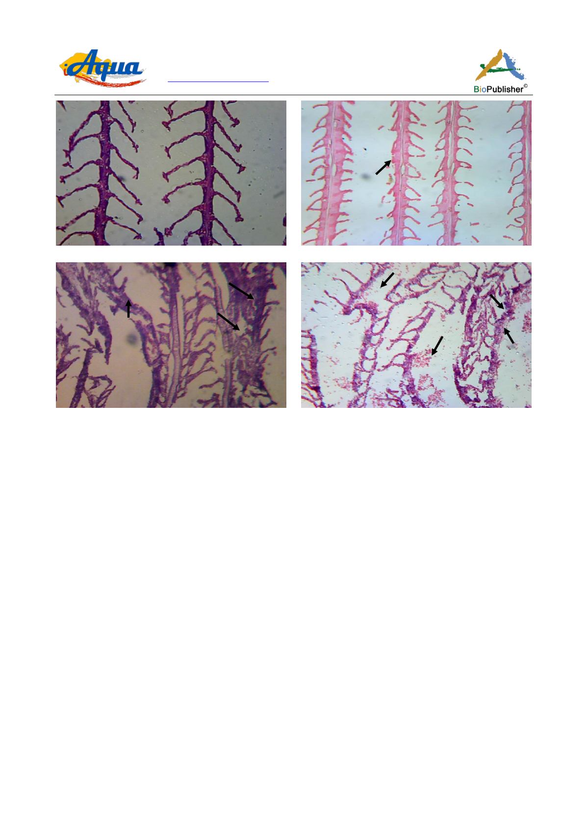
Figure 4 Photomicrograph of gills of
Trichogaster fasciata
after 45 days exposure to chlorpyrifos
Note: A) Control showed normal structure of gills, H&E ×120; B) Hypertrophy (Hy) appeared when exposed to 15 µg/L, H&E ×
150; C) Hemorrhage (H), vacuums (V), missing of gill lamellae appeared when exposed to 50 µg/L, H&E ×125; D) Hemorrhage (H),
vacuums (V), necrosis were observed when exposed to 150 µg/L, H&E ×125
3 Discussion
Histopathological observation is a sensitive bio-monitoring tool in toxicant impact assessment to indicate the
effect of toxicants on fish in pesticides polluted aquatic ecosystems (Marchand et al., 2009). Pesticides in polluted
aquatic ecosystem are accumulated mainly in the metabolically active tissues of fish such as liver, kidney, gonads
and gill (Oruce and Usta, 2007) and cause histopathological damage of those organs.
Trichogaster fasciata
was
exposed to different sub-lethal concentrations. Four sub-lethal concentrations (15, 50, 150 and 500 μg/L) of
chlorpyrifos based on previously estimated LC
50
of 880 μg/L were used in this study for 75 days.
The histopathological changes in gills of common carp (
C. carpio
) exposed to organophosphate pesticide,
malathion at 1.5 and 3.0 ppm was observed by Sharmin (2014). Several morphological changes were seen in the
gills of fish exposed to malathion. The gills of fish exposed to low dose (1.5 ppm) showed Telangiectasia and
Blood Lamellar congestion while Telangiectasia, Blood Lamellar Congestion, Hypertrophy of filaments, Lamellar
Fusion observed in the gills of fish exposed to high concentration (3.0 ppm).
However, at the higher tested concentrations of chlorpyrifos 20 EC viz., 50 and 150 μg/L marked degenerative
changes, severe necrosis, pyknosis, haemorrhage and vacuolation were observed in all the tested fish species
which agreed with the finding of Rahman et al. (2002) and Omitoyin et al. (1999). All of the fishes died within 5
days when exposed to 500 μg/L.
Navaraj and Yasmin (2012) investigated the impact of tannery industry waste water on
O. mossambicus
. The
histopathological changes observed in gills, liver, kidney and brain of the studied fish have demonstrated the
quality status of industrial wastewater. In the vital organs, the following marked changes were observed: filament
cell proliferation, cellular infiltration, haemorrhage and epithelial lifting in gills, vaculation of hepatocytes and
A
B
C
D
Hy
H
V
V
H
H
N


