International Journal of Aquaculture, 2018, Vol.8, No.14, 104-111
109
necrosis in liver, exfoliation and swollen with pyknotic nuclei in kidney and enlarged pyramidal cells, binucleated
nuclei, vaculation, and necrosis in brain.
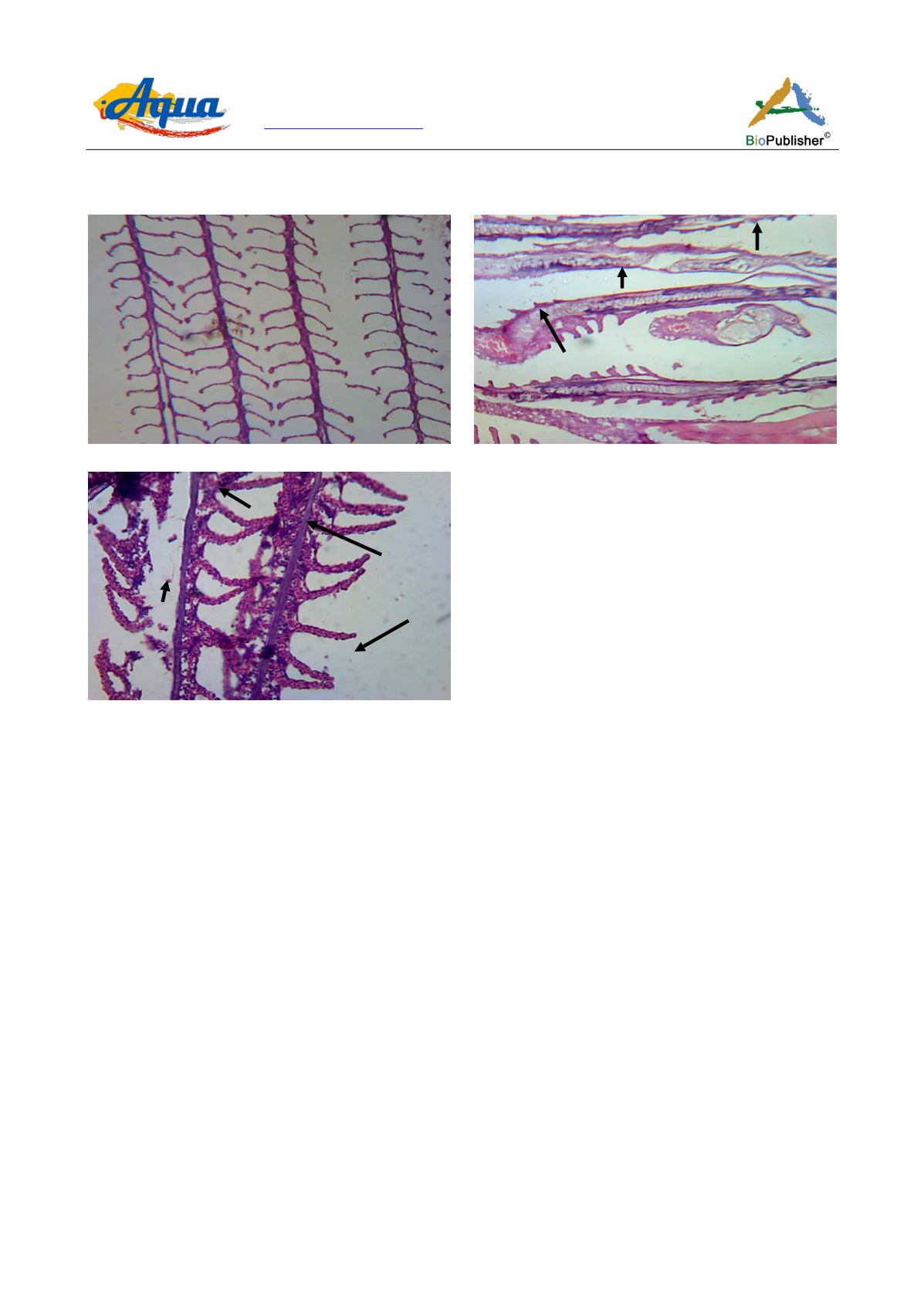
Figure 5 Photomicrograph of gills of
Trichogaster fasciata
after 60 days exposure to chlorpyrifos
Note: A) Control showed normal structure of gills; B) Hemorrhage (H), Hypertrophy (Hy), missing of lamellae, appeared when
exposed to 50 μg/L; C) Pyknotic cells (P), hemorrhage (H), hypertrophy (Hy), splitted gill lamellae (Sgl) were observed when
exposed to 150 μg/L, H&E ×120 magnification was maintained
To determine the histopathological effects of gammalin 20 on African catfish (
C. gariepinus
) an experiment was
conducted by Lawrence and Tamiotan (2010). The 96 hrs lethal concentration (LC
50
) value was 30 ppm.
Histopathological changes of the gill, liver, and intestinal tissues of fish treated with sub lethal concentrations of
gammalin 20 for twelve weeks showed gill distortion and fusion of adjacent secondary lamella as a result of
hyperplasia and excessive mucus accumulation. The liver showed swelling of hepatocytes with mild necrosis,
pyknosis, and vacuolation, while the intestine showed yellow bodies of the lamina propria at the tip of the
mucosal fold.
Degenerative changes in gill, such as intraepithelial edema in the secondary lamellae, thick coating of mucus
covering the entire gill filaments and lamellae, erosion of secondary lamellae, thickening of lamellae,
inflammation of epithelial cells, breakages in primary lamellae, degeneration of secondary lamellae, necrosis,
rupture of epithelium were noticed during exposure of sublethal concentrations of monocrotophos by Rao et al.
(2005). Histopathological changes observed were hemorrhage in the primary and secondary gill lamellae,
degeneration and necrosis of epithelial cells, distortion of the secondary lamellae, disruption of epithelial cells
from pillar cells, shorter gill lamellae, fusion, complete destruction of lamella, increased vacuolation, irregular
appearance of gill lamellae were observed in guppy
Poecilia reticulate
exposed to chlorpyrifos (De Silva and
Samayawardhena, 2002).
However, the result of the present study revealed that chlorpyrifos 20 EC is toxic to fish and causes
histopathological changes in fish organs. The LC
50
values recorded in this study were very lower which indicated
A
B
C
Hy
Sgl
H
Hy
P


