International Journal of Aquaculture, 2018, Vol.8, No.14, 104-111
107
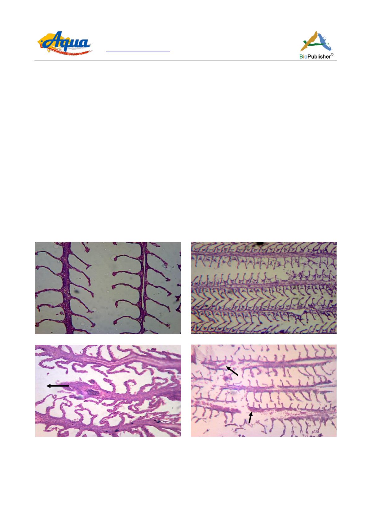
concentration. Notable structural changes occurred at a concentration of chlorpyrifos 150 μg/L like necrosis,
missing of gill lamellae. Chlorpyrifos induced alterations at 45 days in histoarchitecture of the gills were very
significant (Figure 4). Control showed normal structure but changes like hypertrophy, hemorrhage, vacuums and
necrosis were observed after exposure at different concentration of chlorpyrifos (15 μg/L, 50 μg/L, 150 μg/L). The
96-h LC
50
of chlorpyrifos for this species was found to be 880 µg/L.
Greater damage of gills was observed after 60 days exposure of chlorpyrifos like the cells of gills got thickness
and were reduced into a dense solid mass (pyknosis), increased in size of cells (hypertrophy), escaped of blood
from blood vessels and occurred in the membrane, skin and vessels (hemorrhage), circumscribed death of cells
with black structural evidence (necrosis) at different concentrations of chlorpyrifos (Figure 5).
A notable observation with respect to gills damage was observed after 75 days exposure of chlorpyrifos was that,
the gills exposed to the 15 µg/L, 50 µg/L, 150 µg/L concentrations exhibited greater distortion of gills
architechtecture than the 15, 45 and 60 days exposure. Concentration 500 μg/L of chlorpyrifos showed lethal
effect to
Trichogaster fasciata
(Figure 6).
All the histopathogical observations indicated that exposure to sublethal concentrations of chlorpyrifos caused
destructive effect in the gills of
Trichogaster fasciata
. Gill histopathogical alterations, such as those observed in
this study could result in severe physiological problems which ultimately leads to the death of fish. The findings
of the present histological investigations demonstrated a direct correlation between chlorpyrifos exposure and
histopathological disorders observed in gills.
Figure 3 Photomicrograph of gills of
Trichogaster fasciata
after 15 days exposure to chlorpyrifos
Note: A) Start of the experiment, H&E × 120; B) Almost normal structure appeared when exposed to 15 µg/L, H&E × 150;
C) Hypertrophy (Hy) appeared when exposed to 50 µg/L, H&E ×120; D) Necrosis (N), missing of gill lamellae were observed when
exposed to 150 µg/L, H&E ×125
A
B
C
D
Hy
N


