Computational Molecular Biology 2016, Vol.6, No.1, 1-20
8
Note: (a) Domain architecture showing an N-terminal RING domain (green) preceded by four zinc fingers (yellow) and TRAF
domain (purple) at the C-terminus. (b) ribbon representation showing RING domain of TRAF6 (green) binding to E2 enzyme (PDB
ID: 3HCT). The first zinc finger (Z1) along with the RING domain is involved in TRAF6-E2 interaction. Grey balls represent zinc
ion.
4.1.2.2 Parkin
Misfolded protein misfolding and aggregation is a hallmark of the neurodegenerative disorder, Parkinson’s (Tan et
al., 2009). Malfunctioning UPS system contributes to the pathological progression of the disease (Lim and Tan,
2007). Mutations in parkin, an RING E3 ligase contributes to around 50% of the cases of juvenile parkinson’s.
Parkin is involved in maintaining mitochondrial quality and mitochondrial autophagy (Figure 3). Upon signal
reception, parkin ubiquitinates mitochondrial membrane proteins leading to their degradation and hence
elimination of any damaged organelle. Mutations in parkin gene lead to accumulation of damaged mitochondria
which is a source of reactive oxygen species (ROS). ROS is a major cause of neural cell degeneration (Durcan and
Fon, 2015; Tanaka, 2010). Parkin belongs to the unique RING-between-RING (RBR) sub-class of RING E3
ligases. The RBR type of ligases is known to have a active site Cys residue in addition to the usual RING domain.
Hence, they function as RING as well as HECT type E3 ligases. Structure of parkin involves an ubiquitin (Ubl)
domain in its N-terminal and RBR domain in the C-terminal end. The overall structure of RBR domain consists of
two compact domains. First has the merged R1 and in-between R (IBR) domain structure. R1 domain represents
the classical RING domain cross-brace structure and is known binds E2. Second domain comprises the R0 and R2
in close proximity. The R2 domain comprises of the catalytic Cys 431 residue. Mutation of the hydrophobic
residues in R0-R2 domain results in autoubiquitination of parkin. The IBR domain forms a bi-lobed structure and
co-ordinates two Zn ions. Mutations in particular regions leads to the malfunctioning parkin : 1) In the Zn binding
residues of the R1 domain resulting is structure collapse 2) residues binding to E2, inhibiting the E3-E2 intractions
3) in the active site pocket with Cys 431(Beasley et al., 2007; Riley et al., 2013). As loss-of function mutations in
parkin gene leads to the onset of Parkinson’s syndrome, drugs inhibiting the autoubiquitination of parkin may
prove to be neuroprotective.
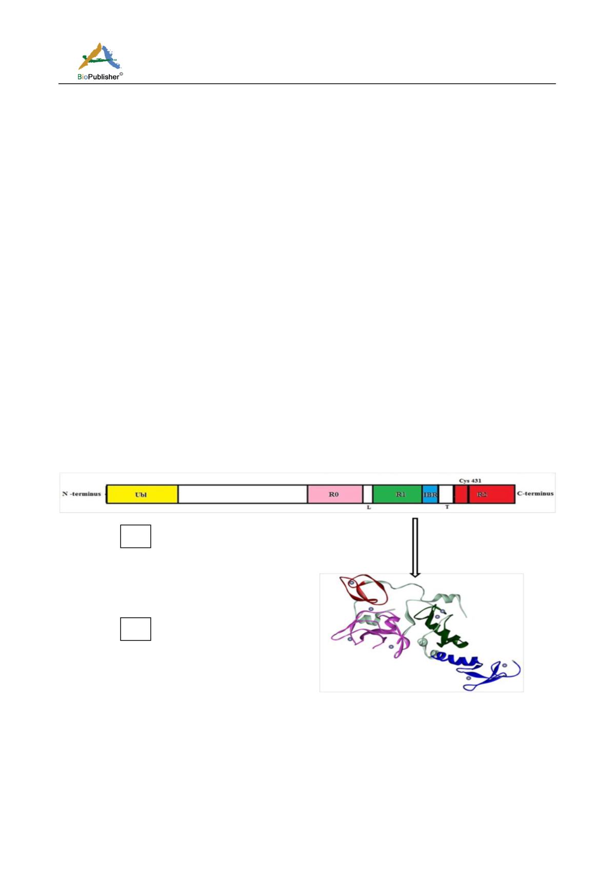
Figure 3 Parkin
Note: (a) Domain architecture of parkin (RBR) E3 ligase. Ubl (ubiquitin binding domain) in N-terminus, yellow binds E2 enzyme.
RBR domain is located at the C-terminus consists of R0(pink), R1(green), IBR (blue) and R2(red). R1 represents t he real criss-brace
RING domain, while the R2 consists of the catalytic Cys 431 residue and act as a HECT domain. (b) Ribbon representation of parkin
protein (PDB ID: 4I1F) showing R2(red) in close proximity to R0 (pink). L and T means linker and tether respectively. Grey balls
represent zinc ion.
(b)
(a)


