International Journal of Clinical Case Reports 2017, Vol.7, No.17, 73-80
75
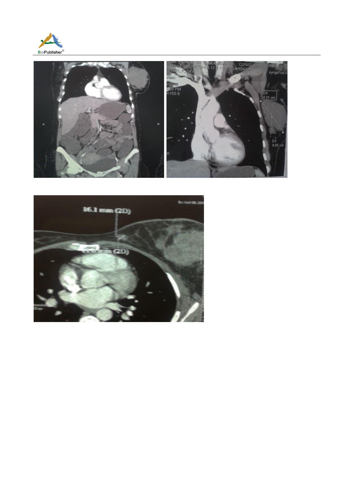
Figure 3
Thoracoabdominal CT: Multiple oval subcutaneous masses of heterogeneous density in left Axillaries region
Figure 4
Thoracic CT: ovarian subcutaneous mass of heterogeneous density in the left anterior thoracic region
The patient was lost sight of despite repeated phone calls. She consulted after 5 months for multiple masses of
lower and upper limbs with increased volume of the left breast.
On examination: appearance of multiple masses of firm, subcutaneous and painless consistency in the lower and
upper limbs, the largest of which sits at the level of the posterior surface of the right thigh and at the level of the
internal surface of the left thigh (Figure 6).
An MRI of both thighs cuts was requested axial and coronal; we note the presence of a bulky tumor mass
developing at the level of the posterior compartment of the lower end of the right thigh, It is in isosignal T1 with
respect to the muscle, in hyper signal T2 with intense enhancement and finely heterogeneous after gadolinium
injection, this mass pushes back the superficial femoral vascular axis which remains permeable with no sign of
bone infiltration (Figure 7).
The patient was placed on Doxorubicin (adriamycin) monotherapy which is the reference treatment. There was a
good clinical response to 5 courses of chemotherapy with decreased size of all clinical targets, we did not opt for
intensive chemotherapy. The course of action was to undertake maintenance chemotherapy with (navelbine)


