International Journal of Clinical Case Reports 2017, Vol.7, No.17, 73-80
77
right lumbar masses) (Figure 8) and also the appearance of a basal posteral pleural nodule that measures 12 mm
with a stable appearance of other lesions (Figure 9).
The progression was marked by the aggravation of pain and the appearance of other tumor lesions in the upper
and lower limbs. The patient died in an acute respiratory distress chart most likely related to pulmonary embolism.
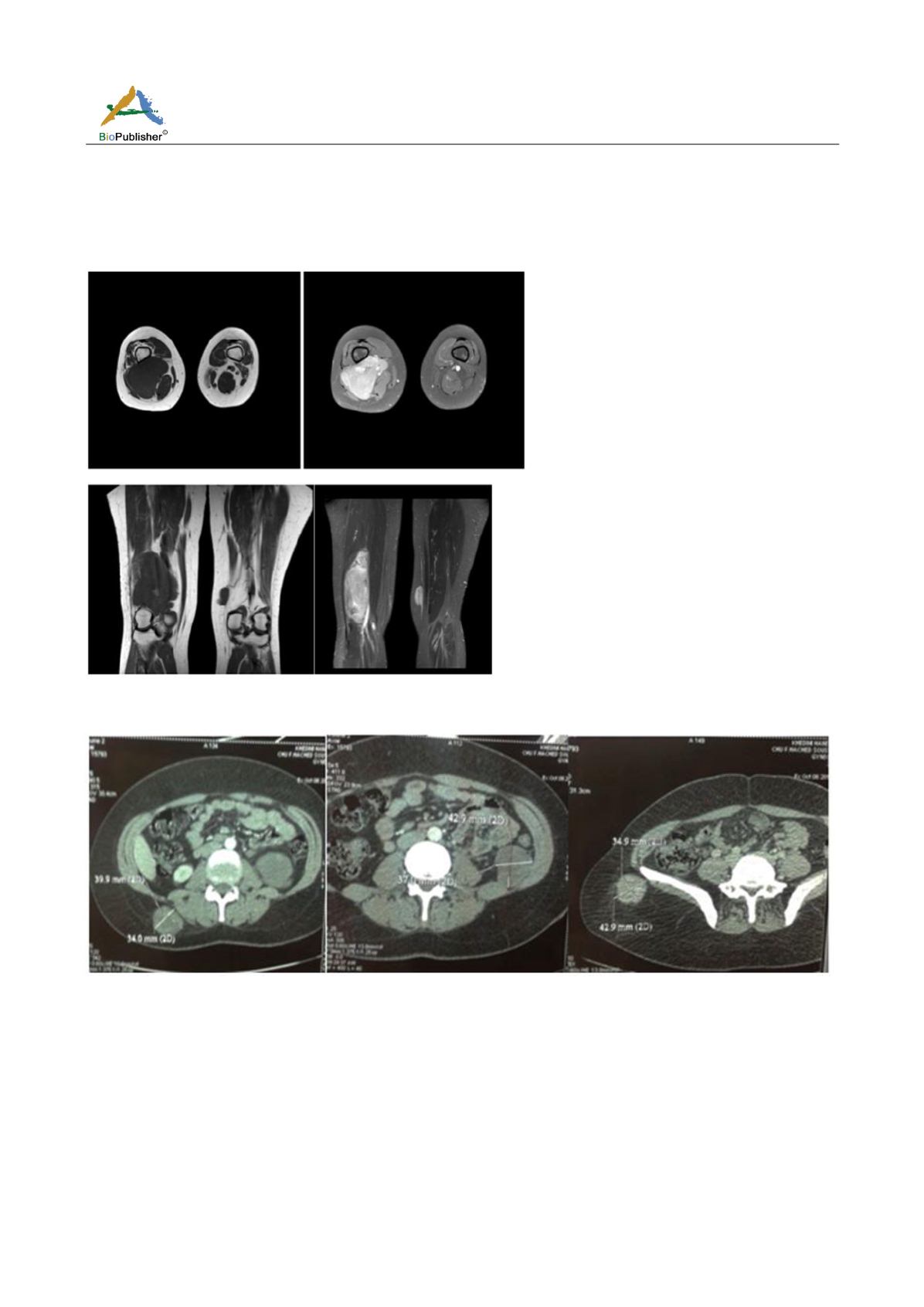
Figure 7 An MRI of both thighs cuts axial and coronal; we note the presence of a bulky tumor mass developing at the level of the
posterior compartment of the lower end of the right thigh
Figure 8 Pelvic abdominal CT: appearance abdominopelvic and parietal lesions (gluteal and right lumbar masses)
2 Discussion
Our patient presented multiple synchronous locations and she had as a circumstance of discovery a breast nodule.
The relevance of our case is in the clinical presentation of the disease and in the particular type of liposarcoma in
its multifocal form.
Soft Tissue Multifocal Liposarcoma is a rare entity in pathology and accounts for only 1 percent of adult
malignancies. Liposarcoma is characterized as multifocal when it appears simultaneously in two or more locations,
before extension to areas where it is more frequently metastasized, such as lung, liver and bone.


