International Journal of Clinical Case Reports 2017, Vol.7, No.17, 73-80
74
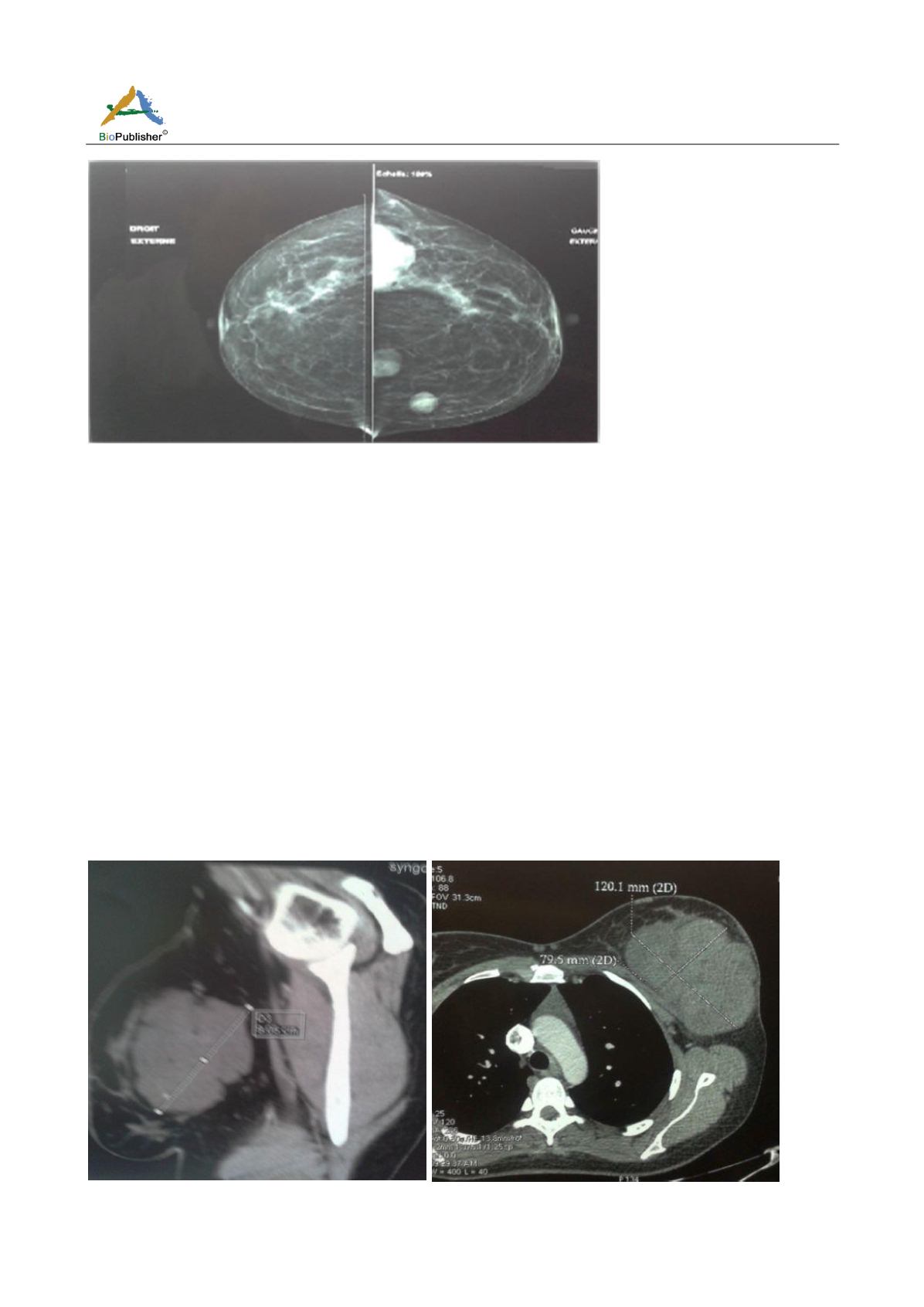
Figure 1
Mammography of both breasts
The patient complained of persistent mastodynia, with a sensation of increased size of nodules palpated a month
after echomammography.
In view of the persistence of mastodynia, a mammary micro-biopsy of the masses was performed and concluded a
morphological appearance and immunohistochemical profile of a myxoid liposarcoma.
The patient reported subcutaneous masses in the upper and lower limbs, of soft and non-painful consistency that
were neglected. An ultrasound of the soft parts was made and concluded to lipomatous masses of
well-circumscribed soft parts, without signs of muscular or bone invasion.
Despite the reassuring ultrasound appearance of these masses, an excisional biopsy was made of one of the masses
at the level of the forearm and concluded that a malignant tumor of high grade of probably sarcomatous nature
recall the appearance of a liposarcoma myxoid.
Thoraco-abdominopelvic CT was performed as part of the myxoid liposarcoma extension assessment and
concluded that multiple tissue masses were present in the left breast (Figure 2), left subcutaneous axillary (Figure
3), thoracic parietal (Figure 4), left gluteal, lumbar and intra and retro peritoneal (Figure 5); They were of the
same finely heterogeneous nature before and after injection of contrast product and exerting a mass effect on
neighboring structures without evidence of obvious invasion.
Figure 2
CT Thoracic: Multilocular subcutaneous oval mass of heterogeneous density of the left breast measuring 12*8 cm


