International Journal of Clinical Case Reports 2015, Vol.5, No. 42, 1-6
2
Case Report
A 23 year old male patient reported the Department of
Oral Medicine and Radiology with a chief complaint
of swelling in lower front tooth region and face since
last 1 year when he suddenly noticed a small swelling
in the lower right front tooth region inside the mouth
and he took some medication. After taking those
medications, the swelling completely subsided. After
6 months, similar swelling appeared which was
progressively increasing in size and was not subsided
by taking medication. The swelling continued
increasing in size and affected the other teeth on
opposite side also. After 10 days, he noticed swelling
on face in mirror. He immediately went to local
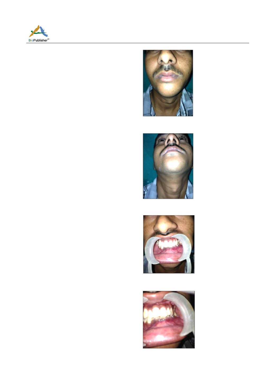
dentist who advised for a biopsy. Clinical examination
revealed a single, ill defined swelling present in
relation to the lower 1/3 rd of the face near symphysis
and parasymphysis regions which crossed midline and
was measuring approximately 5 X 3 cm in diameter
extends mesiodistally from right parasymphyseal
region to left parasymphyseal region and superioin-
feriorly from the vermilion border of lip of right side
and corner of mouth of left side till 1 cm below the
lower border of mandible. Swelling appeared to be
with smooth surface with shiny, slightly stretched skin
and on palpation, swelling appeared to be bony hard
in consistency with well-defined margins and with a
smooth lobular surface (Figure1). Swelling was slightly
tender on right side with no local rise in temperature.
Superficial skin was pinchable with no secondary
changes (Figure 2). Intra-orally, mandibular vestibule
was obliterated by swelling in relation to 35 to 43
region. On detailed intraoral examination, a single
well defined swelling was present in anterior part of
mandible extending from 35 to 43 region with
buccolingual expansion. Buccally, swelling extended
mesiodistally from mesial aspect of 43 to the distal
aspect of 35 crossing midline and buccolingually from
the alveolar crest to the buccal and labial vestibule
obliterating the vestibular region. Superficial alveolar
mucosa appeared to be stretched with detachment of
the marginal gingiva in relation to 33, 34, 35. Swelling
was slightly tender on right side. Swelling was bony
hard in consistency with egg shell crackling which
was present distal to 33. Bucco-lingual expansion was
seen evident throughout the swelling (Figure 3, 4). On
pulp vitality test, 32 was found to be completely
non-responding while 35 showed delayed response.
Figure 1
Figure 2
Figure 3
Figure 4


