Basic HTML Version


International Journal of Marine Science 2014, Vol.4, No.52, 1-9
http://ijms.biopublisher.ca
4
slides were fixed in methanol for ten minutes and
stained with 4% Giemsa solution in phosphate buffer
(pH 6.8). The stained slides were examined under the
light microscope (Labomed Vision 2000, Mumbai,
India) at a final magnification of 1000x. In each slide,
2000 cells with intact cytoplasm were scored
(Barsiene et al
.,
2004; Bolognesi and Fenech, 2012).
The frequency of micronuclei and other nuclear
abnormalities were expressed as mean frequency of
MN/nuclear abnormalities per 1000 cells scored.
The blind scoring of micronuclei and other nuclear
abnormalities was performed on coded slides without
knowledge of the origin of samples. Only cells with
intact cellular and nuclear membrane were scored.
Round or ovoid-shaped non-refractory particles with
colour and structure similar to chromatin with a
diameter 1/3
rd
of the main nucleus and clearly
detached from it were interpreted as micronuclei. In
general, colour intensity of MN should be the same or
less than that of the main nuclei.
1.4 Statistical Analysis
The LC
50
values and 95% confidence interval
endpoints of
M. philippinarum
exposed to Cu, Cd, Pb,
Zn and Hg were calculated using Probit analysis
computer software program (USEPA, 1994). Mean
and standard error was calculated for each experimental
group using Microsoft excel. Non-parametric Dunnett
test was used to compare MN and other nuclear
abnormalities frequencies between control and
treatment groups (P < 0.05).
2 Results
In the present study, the median lethal concentrations
and induction of micronuclei and binuclei were
calculated using
M. philippinarum
exposed to acute
concentrations of copper, cadmium, lead, zinc and
mercury using continuous flow through bioassay test
method.
2.1 Acute Toxicity
The results of the present study showed that the
mortality of mollusc,
M. philippinarum
exposed to Cu,
Cd, Pb, Zn and Hg was increased with increasing
concentration of metal as well as exposure duration.
However, 100% mortality was not noticed in the
highest concentration of all five metals. No mortality
was observed in animals from control (seawater)
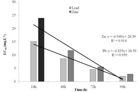
group. The calculated LC
50
values for
M. philippinarum
decreased with increasing exposure period (Figures 1a
and 1b). The 96 h calculated LC
50
values were 0.019
mg. L
-1
Cu [confident interval (CI) values: 0.010 –
0.029]; 0.158 mg. L
-1
Cd (CI values: 0.015 – 0.383);
2.025 mg. L
-1
Pb (CI values: 0.126-4.558); 2.823 mg.
L
-1
Zn (CI values: 1.962 – 7.104) and 0.007 mg. L
-1
Hg (CI values: 0.002 – 0.022). The above results
reveal that Hg was highly toxic and Zn was least toxic
to
M. philippinarum
.
Figures 1a Toxicity for
M. philippinarum
exposed to Cu, Cd
and Hg under acute continuous flow-through bioassay tests
Figure 1b Toxicity for
M. philippinarum
exposed to Pb and Zn
under acute continuous flow-through bioassay tests
2.2 Induction of Micronuclei (MN)
In control groups, the observed mean MN frequency
was the range between 0.3 and 0.5 while the mean
MN frequency calculated was 12.0, 12.7, 5.7, 6.3 and
14.7 in the maximum acute concentration of copper
(0.16 mg. L
-1
), cadmium (14.864 mg. L
-1
), lead (0.96
mg. L
-1
), zinc (16.444 mg. L
-1
) and mercury (0.034
100
counted
cells
of
number
Total
BN or MN
containing
cells
of
Number
BN %or MN%

