International Journal of Aquaculture, 2017, Vol.7, No.1, 1
-
8
5
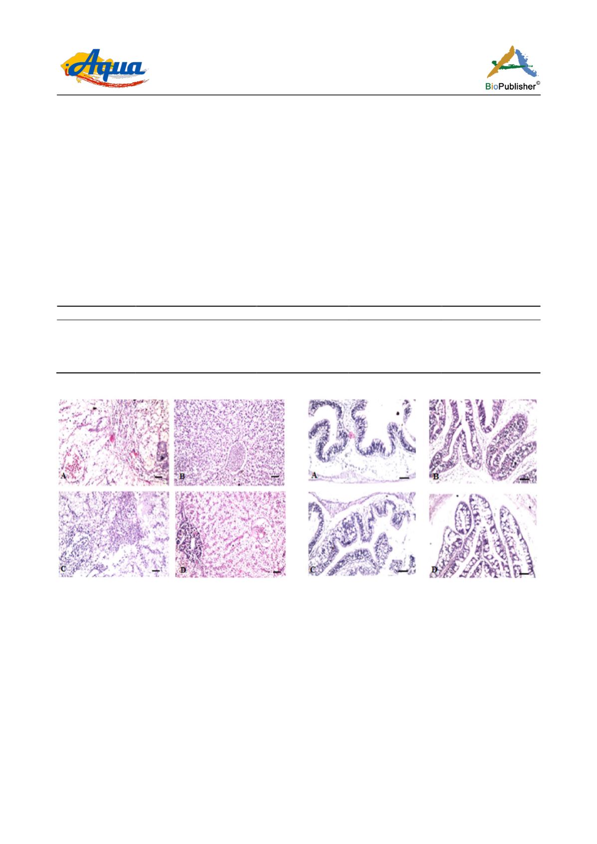
dilatation and congestion were detected in the central veins in the liver structure (Figure 2 B). Also, the hepatic
parenchyma showed focal haemorrage as well as focal inflammatory cells infiltration of fish fed diet
supplemented with moringa leaves meal at 4 g kg
-1
(Figure 2 C). Additionally, dilatation was detected in the
central vein and sinusoids of fish fed diet supplemented with moringa leaves meal at 6 g kg
-1
for 12 weeks (Figure
2 D). Normal structure was observed in the intestine of fish fed the control diet without any supplementation
(Figure 3 A). However, the intestine of fish fed diets supplemented with moringa leaves meal at different levels (2,
4 and 6 g kg
-1
) showed diffuse goblet cells formation in the lining mucosal epithelium associated with
inflammatory cells infiltration in the underlying lamina propria (Figure 3 B, Figure 3 C, and Figure 3 D). Also,
normal structure was observed in the ovary of fish fed the control diet without any supplementation (Figure 4 A).
There were few primary follicles and multiple mature one in the parenchyma. Also, the same results were
observed in the ovary of fish fed diets with different levels of moringa leaves meal (Figure 4 B, Figure 4 C, and
Figure 4 D).
Table 4 The effect of moringa leaves meal as feed additive in the diets on blood components of juvenile Nile tilapia,
Oreochromis
niloticus
(initial wt 2.1 ±1 g) for 12 weeks, Values are mean ±SD of triplicate groups
Treatments
Total protein
TP (g/dl)
Glucose
(mg/dl)
Cholesterol
mg/dl)
Triglycerides
mg/dl)
Control
8.60 ±0.1
a
61.3 ±1.5
d
41.1 ±1.2
a
55.3 ±1.5
b
2 gkg
-1
8.10 ±0.1
a
53.7 ±1.5
c
66.2 ±1.0
c
61.1 ±1.3
c
4 gkg
-1
6.20 ±0.1
b
35.3 ±1.5
a
52.1 ±1.1
b
61.2 ±1.1
c
6 gkg
-1
9.30 ±0.0
a
51.0 ±1.0
b
51.7 ±1.2
b
52.4 ±1.0
a
Note: Means in the same column with different letters are significantly different at (P < 0.05)
Figure 2 Histopathological changes in liver of Nile tilapia fed
different diets (A, Control) without any supplementation,
followed by three diets supplemented with moringa leaves meal
at 2, 4 and 6 g kg
-1
diet (B, C, D, respectively). (A) Showing
congestion was observed in the central veins associated with
focal inflammatory cells infiltration in the hepatic parenchyma.
(B) Dilatation and congestion were detected in the central veins
in the liver structure of fish fed diet supplemented with
moringa leaves meal at 2 g kg
-1
. The hepatic parenchyma
showed focal haemorrage as well as focal inflammatory cells
infiltration (C) of fish fed diet supplemented with moringa
leaves meal at 4 g kg
-1
.There was dilatation in the central vein
(D) of fish fed diet supplemented with moringa leaves meal at 6
g kg
-1
.(H&E; staining) scale bars = 40 µm
Figure 3 Intestine histology of Nile tilapia fed different diets (A,
control) without any supplementation , followed by three diets
supplemented with moringa leaves meal at 2, 4, 6 g kg
-1
diet (B,
C, D, respectively) for 12 weeks. (A) The intestine of fish fed
control diet showed few inflammatory cells infiltration in the
mucosal layer. Diets supplemented with moringa leaves meal
(B, C, D) at different levels exhibit diffuse goblet cells
formation was observed in the lining mucosal epithelium
associated with inflammatory cells infiltration in the underlying
lamina propria. (H&E staining); scale bars = 40 µm


