Cancer Genetics and Epigenetics 2017, Vol.5, No.4, 17-24
18
brain stem compression.
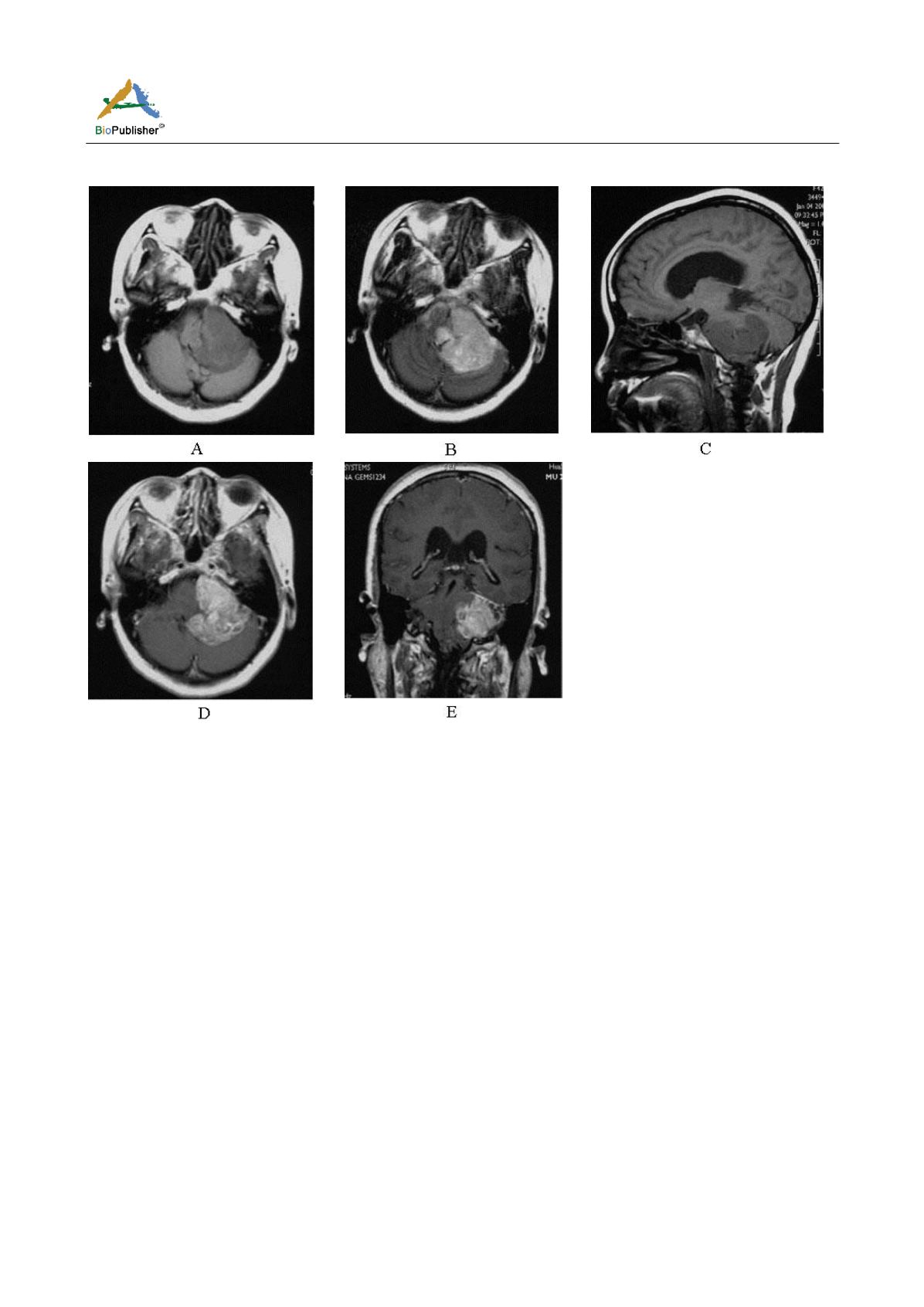
Figure 1 MRI findings of different sequences at axial, sagittal and coronary plane
Note: A: An axial; B: An axial hyperintense lesion on T2-weighted image; C: A sagittal T1-weighted image; D: An axial; E: A
coronary T1-weighted gadolinium-enhanced scan
1.3 Surgical procedure and histopathological findings
Intraoperatively, the reddish lobulated lesion was found to be poor demarcated from cerebellum, and intensely
vascularized. The lesion was seen encasing vestibular and posterior cranial nerves and extending into the fourth
ventricle. Histopathology was performed by 2 independent neuropathologists and revealed that the tumor was
typical of an anaplastic ependymoma (World Health Organization grade III).
2 Discussion
2.1 Pathogenesis
Accounting for approximately 3% of all primary central nervous system neoplasms, ependymal tumors are rare
neoplasms of neuroectodermal origin arising from ependymal cells in the obliterated central canal of the spinal
cord, the filum terminale, choroid plexus or white matter adjacent to the highly angulated ventricular surface
(Patel, 2012; Yang, 2016). Additionally, ependymal tumors can be found in the brain parenchyma as a result of
fetal ependymal cell rests migrating from periventricular areas. One hypothesis regarding oncogenesis of
intracranial extra-axial epedymomas has been proposed and favored by most studies on the basis of the
relationship between the lesion and subarachnoid space: intracranial extra-axial epedymomas derive from ectopic
ependymal nests that result from migration disorders of the germinal matrix. According to the 2016 WHO
classification of CNS neoplasms, ependymal tumors are being classified as WHO grade I: myxopapillary
ependymoma (occurring almost exclusively in the conus-cauda-filum terminale region) and subependymoma, a
benign, slowly growing intraventricular lesion with a very favourable prognosis, WHO grade II ependymoma and
WHO grade III anaplastic ependymoma (Chen, 2017). Although most ependymal tumors are benign, Grade III


