Basic HTML Version


International Journal of Marine Science 2013, Vol.3, No.37, 295-305
http://ijms.sophiapublisher.com
300
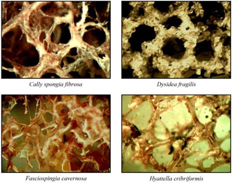
Table 2 Identification and key characteristic features of the four experimental marine Demosponges
Gross Image
Species Name and Classification
Key Characteristic Features
Fasciospongia cavernosa
Phylum: Porifera
Class: Demospongiae
Order: Dictyoceratida
Family: Thorectidae
Massive, keratose sponge with peduncles.
Conulose surface is in dark green to purple
brown and internally grayish yellow. Some
forms resemble
Ircinia species
even in the
symbiotic hostage to many organisms
.
Body
is of fleshy consistency.
Callyspongia (Cladochalina) fibrosa
Phylum: Porifera
Class: Demospongiae
Order: Haplosclerida
Family: Callyspongiidae
Irregular, ramose sponge with cobweb-like
uneven surface appears in brownish purple
to pale yellow. Sub-cylindrical branches
and prominent osculate appear most of the
forms. Body consistency and texture is hard
and brittle.
Hyattella
cribriformis
Phylum: Porifera
Class: Demospongiae
Order: Dictyoceratida
Family: Spongiidae
Growth form tubular, erect and repent. Flat
encrustations of the body, without any proper
shape due to repeated folds. Externally pale
yellow to orange brown or brownish yellow
to green. Body texture is hard and little
compressible.
Dysidea fragilis
Phylum: Porifera
Class: Demospongiae
Order: Dictyoceratida
Family: Dysideidae
Cobweb-like surface formed by huge number
of spicules and sand particles, irregularly
encrusting or massive and lobe-shaped.
Consistency is very brittle to hold and
externally appears pale pinkish to pale
yellow or brownish to grayish-white.
Figure 1 Video micrographs of tangential sections of four
marine sponges
Note: Image magnification at 220× for
F. cavernosa
and at
300×
for
C. fibrosa
,
H. cribriformis
and
D. fragilis
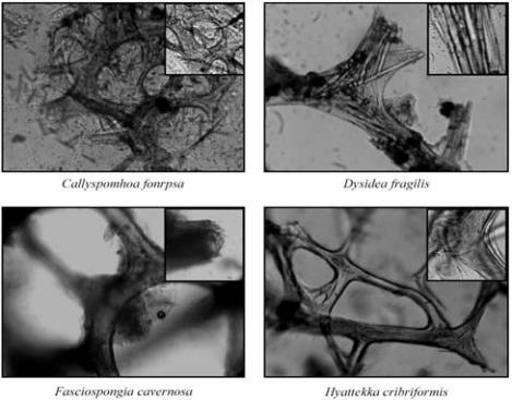
Light Microscopy
The sponge skeletal matrix was observed under light
microscope to evaluate the specular arrangement and
the branching pattern of each sponge species (Figure 2).
Figure 2 Light micrographs of the spongin network of four
experimental sponges (magnification 100×)
Note: Insets representing the high magnification (330×) images
Sponge skeletal network is formed by the intervening
of spongin fibers, spicules and some amorphous
material to give a unique distribution of the spongin
frames. These spongin frames are of different shapes