Basic HTML Version


Int. J. of Marine Science 2012, Vol.2, No.6, 43
-
50
http://ijms.sophiapublisher.com
48
filtered through Whatmann filter paper (No: 2) and
concentrated using vacuum rotary evaporator (Super
fit, Bangalore). The concentrated extract was used for
antimicrobial study. Among the twenty two sponges,
four species were selected for further study based on
the screening tests (Figure 1).
3.2 Antimicrobial activity
For the antimicrobial screening 5 species of bacterial
isolates and three species of fungal isolates were
selected. The bacterial and fungal strains were
obtained from National Collection of Industrial
Microorganisms (NCIM), Pune, India.
Escherichia coli
(NCIM 2065),
Salmonella abony
(NCIM 2257),
Pseudomonas aeruginosa
(NCIM 5031),
(Gram negative bacteria)
Bacillus subtilis
(NCIM
2063),
Staphylococcus aureus
(NCIM 2079), (Gram
positive bacteria) strains were used.
Candida albicans
(NCIM 3102),
Saccharomyces cerevisiae
(NCIM
3054), unicellular fungi and
Aspergillus níger
(NCIM
501) mold fungi were used as fungal test
microorganisms.
3.3 Antibacterial activity of well assay method
Assays were performed according to the standard
guidelines of the National Committee for Clinical
Laboratory Standards (NCCLS, 1999) using a
modified Kirby–Bauer well assay method. A sterile
stainless steel borer (6 mm) was used to make well in
the medium. All the organisms were stored at
-
20
℃
until use. Cells were grown at 3
℃
in Mueller-
Hinton broth to an OD
420
= 1.9 (approximately
10
5
CFU/mL), and were transfer to Muller Hinton
agar for bacteria, and Sabouraud dextrose agar for
yeasts and fungi. Broth cultures were swabbed onto
respective agar medium to achieve a lawn of confluent
microbial growth separately for each strain. Four wells
were bored in each plate. The sponge extract 100 µg/mL
was loaded in to the well and to find out the inhibitory
potential. The plates were incubated for bacteria at 37
℃
24 h and fungi were grown at 28
℃
for 48 h. The
growth of bacteria and fungi around each well was
observed carefully and the diameter of the zone of
inhibition around each well was measured using a
Hi-media zone reader Triplicate plates were
maintained for each test.
3.4 Preliminary screening of sponges for chemical
constituents
Qualitative analysis of the chemicals present was
carried out using methods described by Harborne
(1998). The freshly prepared sponge extracts were
analyzed for the presence of various constituents as
described by Okawori et al (2008) (Table 4).
3.5 Thin Layer Chromatography (TLC)
The sponges with higher antibacterial activities were
taken for TLC studies.
Aurora globostellata
(Carter)
(TCN-8),
Spirastrella
inconstans
var.
moeandrina
Dendy. (TCN-10). Thin layer chromatography plates
were prepared by using with Si-gel F254 grade
(Merck, Darmstadt, Germany) as stationary phase.
Liquid mobile phases were either semipolar (CH
2
Cl
2
:
MeOH; 9:1, v/v) or non polar (Hexane: EtOAc; 8: 2,
v/v). Reversed phase (RP) was used for polar fractions.
The mobile phase systems were MeOH: H
2
O; 3:7, 8:2
and 1:1 (v/v). A one-dimensional ascending
development technique was used to detect the
constituents of an extract on TLC plate. Visual
detection was done in daylight and under UV light at a
wave length of 254 nm. The results were given in
(Figure 3, 4)
3.6 Column Chromatography
In this study, two different sizes of columns were used.
The columns was prepared by using Silica gel as a
packing material which is used as stationary phase.
(Normal phase Column Chromatography) Si-gel 60~120
mesh with a particle size of 0.004~0.063 mm (Merck)
different combinations of organic solvents such as
hexane, ethyl acetate and methanol were used by step
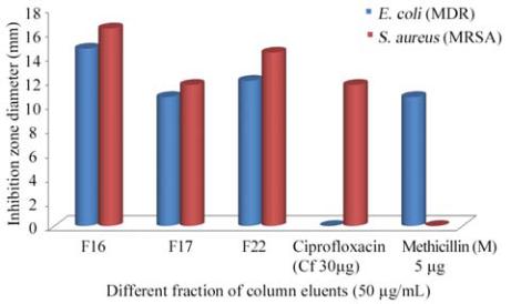
gradient or isocratic elution (Figure 3, 4).
Figure 3 Antibacterial activity of purified compounds from
marine sponge
Spirastrella inconstans
var.
moeandrina
Dendy