International Journal of Horticulture, 2017, Vol.7, No.16, 133-137
136
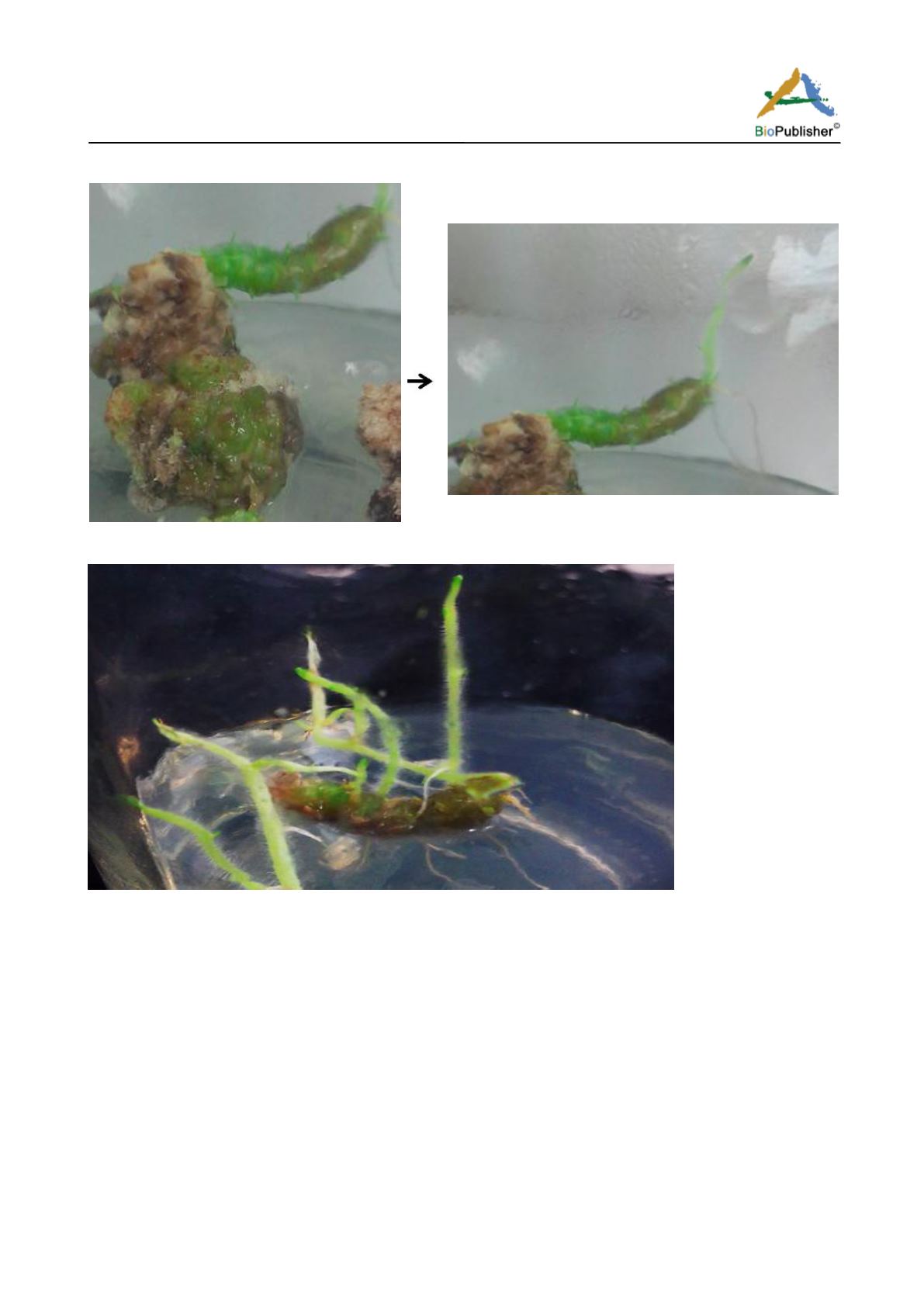
Figure 4 Berries like structure formed on calli shifted to regeneration medium containing 4.75mg/l BAP
Figure 5 Shoot emergence and regeneration from berries like structure
In literature, similar structures were also observed by different scientists but theyall called these, as microtubers.
Kanawl et al. (2006) reported on medium containing 0.75 mg/l BAP, Dhaka and Nailwal (2015) reported on
medium containing 13.18 μM zeatin, 5.71 μM IAA and 8.49 μM GA
3
and Kumar et al. (2014) also reported that
and as well as also stated that these microtubers were sprouted. Hence histoanatomy of these so-called berries
like structure achieved from my research studies were done. Thus, histoanatomy of these so-called berries like
structure and microtubers were performed which revealed that Sclerified layer of cells is visible in epidermal
region in both of the case but in berries it is more prominent because the cells are as less elongated and broad as
twice as long. An indication of lignin deposition is indicated in the epidermal region, as well as inner cortical
region (red coloration) due to Safranin stain, which typically stains secondary wall mainly, composed of lignin
and it is totally absent in microtubers where there is the absence of sclerenchyma in inner cortical parenchyma
(Figure 6; Figure7). In microtubers, numerous starch granules are also visible in the storage region, which is
specific for the tubers (Figure 7). A small constricted area is also indicated in the central portion, which might be
due to collapse of delicate parenchymatous cells during growth pressure which is lacking in so called berries


