International Journal of Clinical Case Reports 2017, Vol.7, No.18, 81-85
82
Ultrasound finds a poorly limited hypoechoic formation og 2.15 cm in diameter with acoustic attenuation but
without doppler vascular flow. The lesion is classified ACR 4 (Figure 2).
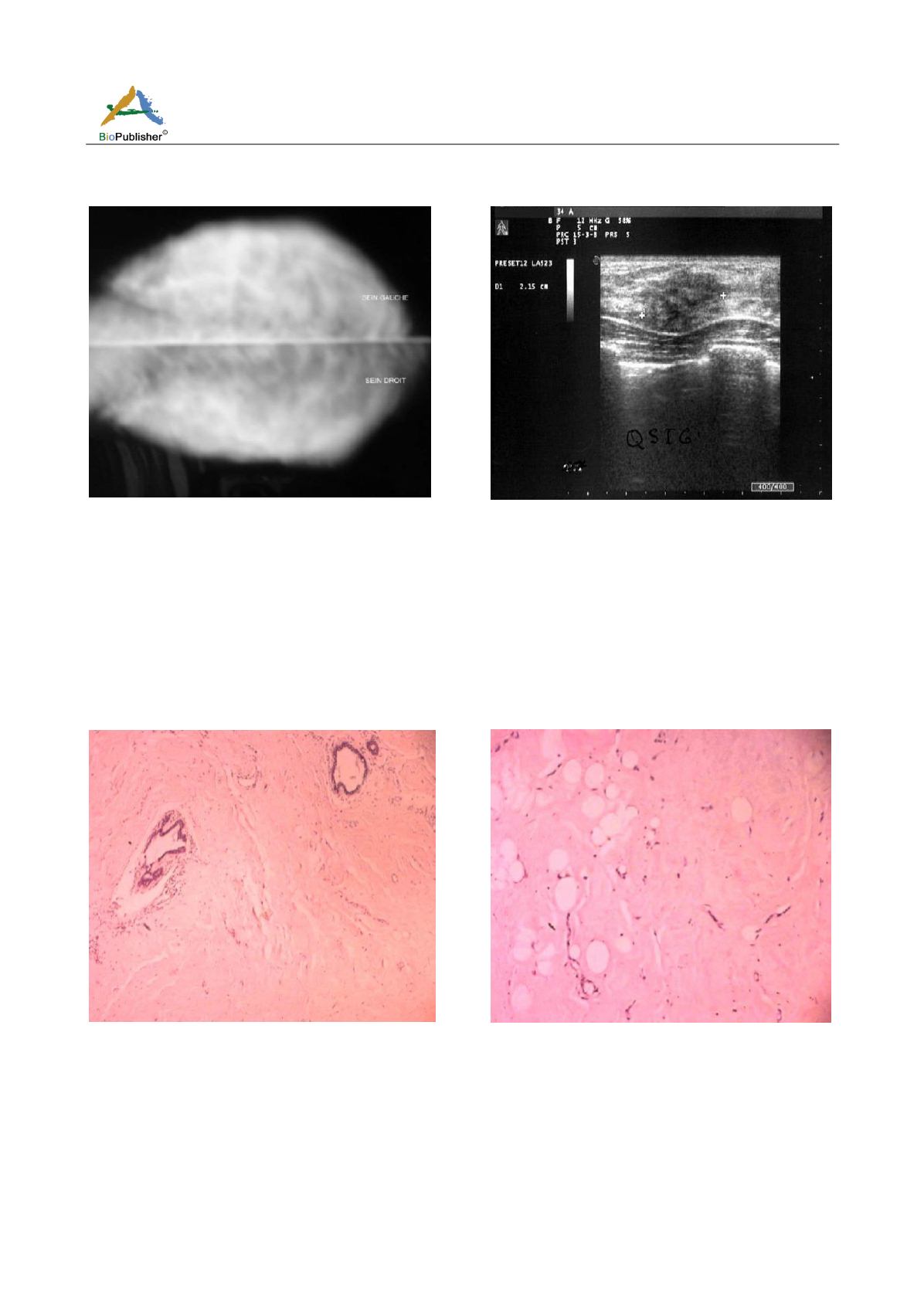
Figure 1 Mammography: denses breasts type IV of BIRADS
without microcalcification and architectural distortion
Figure 2 Ultrasonography: ill-definited margins masses with
marked hypo-echogenicity and posterior acoustic shadowing
measuring 25 cm in diameter
The mammary cytopuncture resulted in a sample insufficient cell with some ductal cells without atypia.
An excisional biopsy of the tumor was performed. The latter is made of hard and dystrophic breast tissue.
Macroscopically the biopsy fragment measures 4 × 3 × 3 cm made of very firm fibrous tissue. Histological
examination showed that the lesion consists of a stroma dense fibrous tissue, with periobular pericanal and
perivascular lymphocyte infiltration. This lymphocyte infiltration is without signs of malignancy, as well as the
presence fusiform fibroblastic cells (Figure 3; Figure 4).
Figure 3 Several lobules has lymphocytic infiltration without
evidence of malignancy
Figure 4 Sclerosing mammary tissu, lymphocytic lobulitis,
which show atrophic changes
Aspect evoking a fibrous mastopathy of the breast. The immediate operative follow-ups are simple, the
medium-term trend was marked by the recurrence of breast nodule at the same location within four month for
which she benefited from a new surgical excision and pathological examination confirmed the fibrous nature of
this mastopathy.


