Medicinal Plant Research 2015, Vol.5, No. 6, 1-9
3
times at 10 and 50 mg/kg As in comparison to
control plants. At 150 mg/kg As, trichome density
decreased by 34%. Trichome density on abaxial epidermis
increased at 10 mg/kg As by 66%, while at 50 and 150
mg/kg As it decreased by 67 and 34 % respectively as
compared to control (Figure. 2).
1.3.2.
Trichome
structure and ultrastructure
LM studies showed that glandular trichomes on the
leaves of control plants were globular, with a stalk of
1-3 cells and a head of four secretory cells (Figure.
3A). At 10 and 50 mg/kg As in soil structural collapse
of the trichome head by disorganization of the
secretory cells and folding of their cell walls was
observed. Trichomes of plants treated with 150 mg/kg As
showed senescence-like symptoms. Head cells showed
mushroom-like appearance, partially protruding out of the
epidermal depression (Figure. 3D). SEM investigations
revealed an equatorial line of weakness around the
head of the trichome in leaves of control plants
(Figure. 4A). The cuticle ruptured along this line, and
the subsequent collapse of the subcuticular cavity led
to the release of exudates. Early maturity of trichomes
was observed in As treated plants. Disintegration of
the secretory cells and folding of the cell walls was
observed in trichomes at 10, 50 and 150 mg/kg As
treatments. Exposed head cells with no cuticle were
noticed at 50 mg/kg As (Figure. 4C). A large number
of deeply grooved trichomes was common at 150
mg/kg As (4D), a typical behaviour of mature
trichomes (Werker, 1993). Wax deposition was more
clearly visible on leaf surface (Figure. 4D). Distortion of
epidermal cells was observed with increased concentration
of As. Non-glandular trichomes also lost the structural
integrity with higher doses of As in soil.
TEM studies provided further insights into the
secretory cells of trichomes. Well-developed central
nucleus and dense cytoplasm was observed in the head
cells of control plants. Large vacuoles and mitochondria
having well developed cristae were seen all over
(Figure. 4E). Rough endoplasmic reticulum (RER)
and dictyosomes were also distinctly visible, signifying
that cells were actively involved in EO secretion. At
10 mg/kg As no significant variation was observed in
head cells in comparison to control. Well developed
nucleus with electron dense chromatin material
was observed in the cells (Figure.4F). However, at
50 mg/kg As, plasmodesmatal connections were
Figure 1. EO yield (%) of
O. basilicum
as affected by different
concentrations of arsenic levels. Values with different
superscripts (a-c) are significantly different at
p
≤ 0.05.
Figure 2. Trichome density (cm
2
) on both adaxial and abaxial
surfaces of
O. basilicum
as affected my different arsenic levels.
Values with different superscripts (a-c) are significantly different at
p
≤ 0.05.
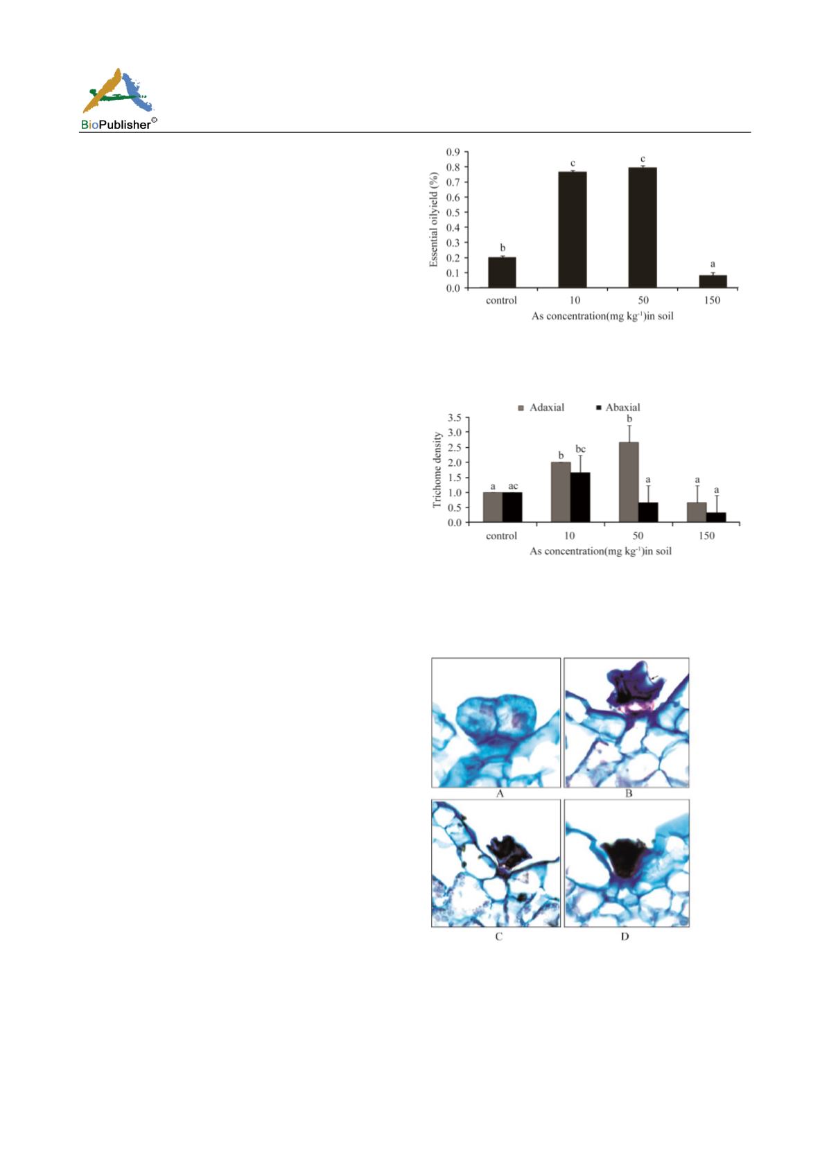
Figure 3. Light micrographs showing the response of O.
basilicum leaf peltate glandular trichomes to arsenic at different
concentrations (40X).
(A)Control: Developing trichome.
(B)10 mg/kg As: Folding of head cells (arrow).
(C)At 50 mg/kgAs: Collapsed head cells and shortening of stalk cell.
(D)At 150 mg/kg As: Senescent trichome.


