International Journal of Clinical Case Reports 2015, Vol.5, No. 41, 1-5
2
parents were advised surgery for the same. Apart from
duodenal atresia, ultrasonogram of abdomen and
pelvis was normal with the liver and spleen and other
organs intact. The parents did not want surgery there
and brought back the child to the local hospital and
from there the baby was referred to our centre.
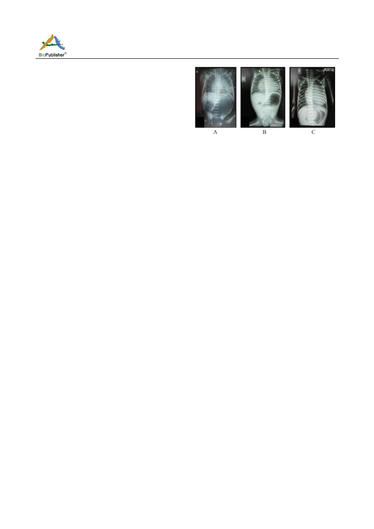
The first Xray taken erect with chest and abdomen
(Figure 1A) at local hospital on day 6 of baby’s life
showed cardiomegaly, pulmonary fields not plethoric,
liver shadow in the right hypochondrium and grossly
distended stomach with a double bubble appeareance.
The baby was admitted for palliative treatment. The
second radiograph (Figure 1B) done here after
admission (after 12 hours after the first Xray) shows
the very clear double bubble appearance. On general
physical examination, baby had stable vitals. He was
afebrile, heart rate 122/minute, respiratory rate 52/minute,
His oxygen saturation was 94% at room air. All
peripheral pulses were equally felt. Blood pressure
was 74/36 mm Hg in right upper limb, 73/35 mm Hg
in left upper limb. 72/76 mm Hg in right lower limb.
76/24 mm Hg in left lower limb. Anterior fontanelle
was normal. There was no central cyanosis and any
significant dysmorphism.His respiratory effort remained
good and was active and at times crying vigourously.
Abdominal examination revealed gross distension of
the abdomen and there was no hepatosplenomegaly.
Cardiovascular system examination revealed S1 normal,
S2 split,an ejection systolic murmur in the left
parasternal border was normal. Respiratory system
examination was normal except for occasionally being
tachypneic. Genito urinary system examination was
normal. He did not have any neurological deficits. The
baby was kept nil orally and hydration with intravenous
fluids and supportive care such as maintenance of
normal temperature, catherisation of urinary bladder
to measure the urinary output, intravenous antibiotics
as per the NICU protocol with Cefotaxim and
Gentamicin was started. The newborn was assessed
by the paediatric surgeon and on day 8 of his life the
Khimora procedure –duodenostomy was done under
local anaesthesia after properly informing the parents
and consent. During the surgery the preportal vein was
found obstructing the distal lumen and there was
grossly dilated stomach and the first part of duodenum.
The baby tolerated the procedure and was in the NICU
for the further care.
Figure 1A Photo of the first Xray taken erect with chest and
abdomen showed cardiomegaly,pulmonary fields not plethoric,
liver shadow in the right hypochondrium and grossly distended
stomach with a double bubble appearance
Figure 1B Photo of the second radiograph shows the very clear
double bubble appearance
Figure 1C Photo of the the third radiograph shows the decompressed
stomach with cardiomegaly and normal lungs
The laboratory investigations were done: On
admission-A quick glucose check was 175 mg/dl.
Hemogram revealed Hb (12.1 g/dl), PCV (33.7%),
total count (11 690/µl), neutrophils (57.6%), lymphocytes
(24.5%), eosinophils (0.3%), monocytes (2.5%), basophils
(0.1%), ESR (31 mm/hr), and platelets count (75
000/µl). Serum Na
+
137 meq/l, K
+
5.1meq/l, Cl
-
87
meq/l, bicarbonate 21 meq/l, glucose 144 mg/dl, total
Ca
++
8.8 mg/dl. C Reactive protein was 19.4 mg/dl
(normal = <0.6md/dl) BT: 2 minutes CT:12 minutes
HIV/HBsAg: Negative, Serum Creatinine: 1.1 mg%,
Serum Ca
++
: 9.0 mg%. Total Serum Bilirubin: 7.7
mg/dl, Indirect serum Bilirubin: 6.3 mg/dl. Urine
routine was normal. On day 4 after admission Serum
Na
+
137 meq/l, K
+
5.1 meq/l, Cl
-
106 meq/l, bicarbonate
16 meq/l, glucose 144 mg/dl, total Ca
++
9.8 mg/dl. C
Reactive protein was 4.7 mg/dl (normal == <0.6 md/dl)
Serum Creatinine: 0.6 mg%, Serum Ca
++
: 9.0 mg%.
Platelet count 100 000 /µl. On day 10 of his life he
had respiratory distress and he became lethargic .His
blood sugars were normal. He was transfused packed
red blood cell concentrate and antibiotics changed to
Meropenam injection and was hydrated properly.
Then his general condition improved. The third
radiograph (Figure 1C) done here after 6 hours after
the second Xray shows the decompressed stomach
with cardiomegaly and normal lungs. But on day 15 of
his life he developed again respiratory distress and on
day 16 of his life (11
th
day after admission) he
suddenly developed desaturation and expired in spite
of all resucitatory measures given. The cause of the


