Genomics and Applied Biology 2015, Vol. 6, No. 7, 1-8
3
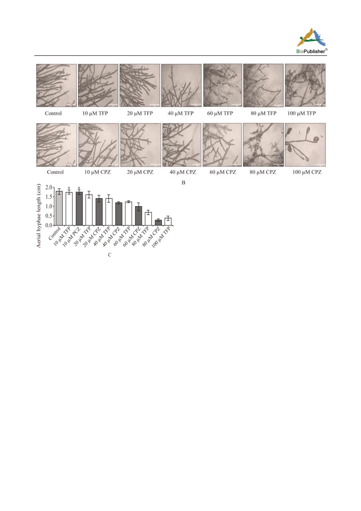
Figure 1 Effect of trifluoperazine (TFP) and chlorpromazine (CPZ)
on growth of
N. crassa
. (A) Effect of TFP and CPZ at various
concentrations on apical growth. (B) Abnormal hyphal
morphology with increasing concentrations of TFP and CPZ. (C)
Aerial hyphae length of cultures grown for 72 h in various
concentrations of TFP and CPZ. Error bars indicate the standard
errors calculated from the data for three independent experiments.
Statistically significant values are indicated by asterisks, *P < 0.05
were verified using polymerase chain reactions (PCR)
of the progeny strains (Supplementary Figure 1).
Growth, crossing, and maintenance of
Neurospora
strains were essentially as described by Davis and De
Serres (1970). The apical growth was analyzed by
using standard race tube assay and calculated as cm
h
-1
(Ryan et al., 1943, 1950). For aerial hyphae, ~1 X
10
6
cells/ml of each strain was grown in liquid Vogel’s
sucrose media (VSM) and incubated at 30
C for 48 h
in dark followed by 24 h light illumination at room
temperature and height of aerial hyphae was measured
(Deka and Tamuli, 2013). Conidial count was done
after 72 hours of growth; a sample of each strain was
withdrawn and harvested using sterile water followed
by conidial counting using a haemocytometer under a
Trinocular Phase Contrast Microscope (Supplementary
Figure 2). For growth yield, ~1 X 10
6
cells/ml of each
strain were inoculated in liquid Vogel’s medium at
30ºC with shaking at 200 rpm for growth. Mycelia
were collected at a regular interval of 24 h by
filtration, dried and weighed over a period 96 h. For
analysis of hyphal morphology, strains were grown for
12 h on a thin layer of Vogel’s agar on glass slide, and
observed under microscope at 20X magnification. In
addition, statistical significance was performed
according to variance analysis (ANOVA, P < 0.05).
Assay for calcium sensitivity and thermotolerance
Assay for calcium sensitivity was done essentially as
described previously (Deka et al., 2011). Briefly,
conidia was placed in the centre of petri dishes
containing Vogel’s glucose (1.5%) media supplemented
with 0.0 M, 0.2 M, 0.3 M, 0.4 M CaCl
2
incubated at
30ºC and colony diameter was measured every 3 h
over a period of 24 h and growth rates were calculated
as cm h
-1
. For measuring thermotolerance, three
days-old conidia were inoculated into liquid Vogel’s
Medium at a concentration of ~1 X 10
6
cells/ml and
germinated for 2 h with shaking at 200 rpm at 30
C.
These germlings were exposed to different heat
treatment condition in two sets one set was held at
30
C for uninduced condition and the other set at
44
C for induced condition for 30 min, then one set of
each were given a lethal heat shock at 52
C for 20 min.
(Yang Qi and Borkovich, 1999; Kumar and Tamuli,
2014). After that these conidia were spread on sorbose
agar (0.05 % fructose, 0.05 % glucose, 2% sorbose, 2 %


