Basic HTML Version


International Journal of Marine Science 2014, Vol.4, No.33
http://ijms.biopublisher.ca
2
Oman, 75 specimens of
L. equulus
ranging in length
65-190 mm SL were obtained and their vertebral
column were studied. To prepare dry vertebral column,
the method of James (2008) was followed. The fish
specimens were boiled to 110˚C to strip the flesh off
the bone. After boiling, their vertebral column was
brushed in running water. After drying, the vertebrae
were separated and numbered then measured with a
digital 1/100 caliper (Ted Pella, Inc., Redding, USA).
The dry vertebrae were deposited in the ichthyological
collection of the marine science and fisheries Centre,
Ministry of Agriculture and Fisheries, Oman, Catalogue
no. 1245. Four vertebral measurements were selected:
vertebral length (VL), the distance along the left
mid-ventral line. It is considered among the factors
controlling the degree of the body’s flexion (Ramzu et
al., 1992); vertebral height (VH) the maximum
vertical distance of the anterior side of the vertebrae;
vertebral width (VW), the maximum horizontal length
across the anterior surface of vertebra; and central
length of vertebra (VCW). The presence of bony
crests might affect significantly the mechanical
constrains exerted by the intermediary of muscles
(Ramzu and Meunier, 1999). The data are presented as
average values (Table 1). To indication any statistical
significant difference is present in the vertebral
measurements,
t
-test was used to show such difference.
From these four measurements, it is possible to
establish a vertebral profile which reflects the
variation of these parameters along the vertebral axis
(Desse et al., 1989, Ramzu, 1994, Kacem et al., 1998,
Ramzu and Meunier, 1999, Nowroozi, 2012). To
avoid individual variation and to facilitate future
comparisons with other samples, even other species,
each vertebral measurement was converted into a
vertebral index V
i
(Ramzu and Meunier, 1999):
V
i
=P/SL
Where, P is the vertebral parameters (VL, VH, VW
and LC) and SL the standard length. Profiles of the
vertebral column were drawn by plotting VL, VH,
VW and LC against the ordinal number of the
vertebrae.
The number of abdominal and caudal vertebrae was
counted and the mean value is calculated for each
vertebra then species means were calculated for
abdominal vertebral number (AVN) and caudal
vertebral number (CVN). The thoracic vertebrae were
defined as those that were cranial to vertebrae with
separated haemal arches. The caudal region was
defined as the region from the first fused haemal arch
posterior to the last centrum including the ural
centrum. The mean vertebral aspect ratio (AR=
centrum height/ centrum width) for each region was
calculated for each individual. The means were then
calculated for abdominal aspect ratio (AAR) and
caudal aspect ratio (CAR). Osteological terminology
mainly follows Chapleau (1988), Ramzu and Meunier
(1999), and Nowroozi (2012).
2 Results
All the twenty three specimens of
L. equulus
analysed
here for gross morphology all had 23 vertebral centra
from cranial to caudal excluding the urostyle: 10
abdominal vertebrae, 13 caudal vertebrae, and the
Urostyle (Figure 1).
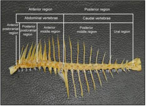
It is possible to divide the
vertebral column of
L. equulus
into five regions:
anterior postcranial region (V1), posterior postcranial
region (V2-V5), anterior middle region (V6-V10),
posterior middle region (V11-V18) and ural region
(V19-V23). The choice to separate the vertebral
column in five regions is supported by differences in
the length and the height of vertebra (
t
≥ 1.98; p ≤ 0.05).
Figure 1 Vertebral column of
Leiognathus equulus
showing
regionalization (Magnification = X1)
Vertebra no. 11 marks the boarder between thoracic
and caudal vertebrae. The 10 vertebrae of the anterior
region, define the abdominal region or truncal,
delimited by the presence of the gut, the two haemal
arches, remain separated and the haemal spine is
absent. The caudal vertebrae belong to the tail; their