International Journal of Marine Science, 2017, Vol.7, No.30, 292-296
294
Normal coloration of the species:
Body mainly with olive green coloration, with a number of indistinct rounded or irregular pale, dark edged areas.
Some of which are with or without a dark central spots. Numerous small dark spots on head, body and fins. The
blind side should be completely white.
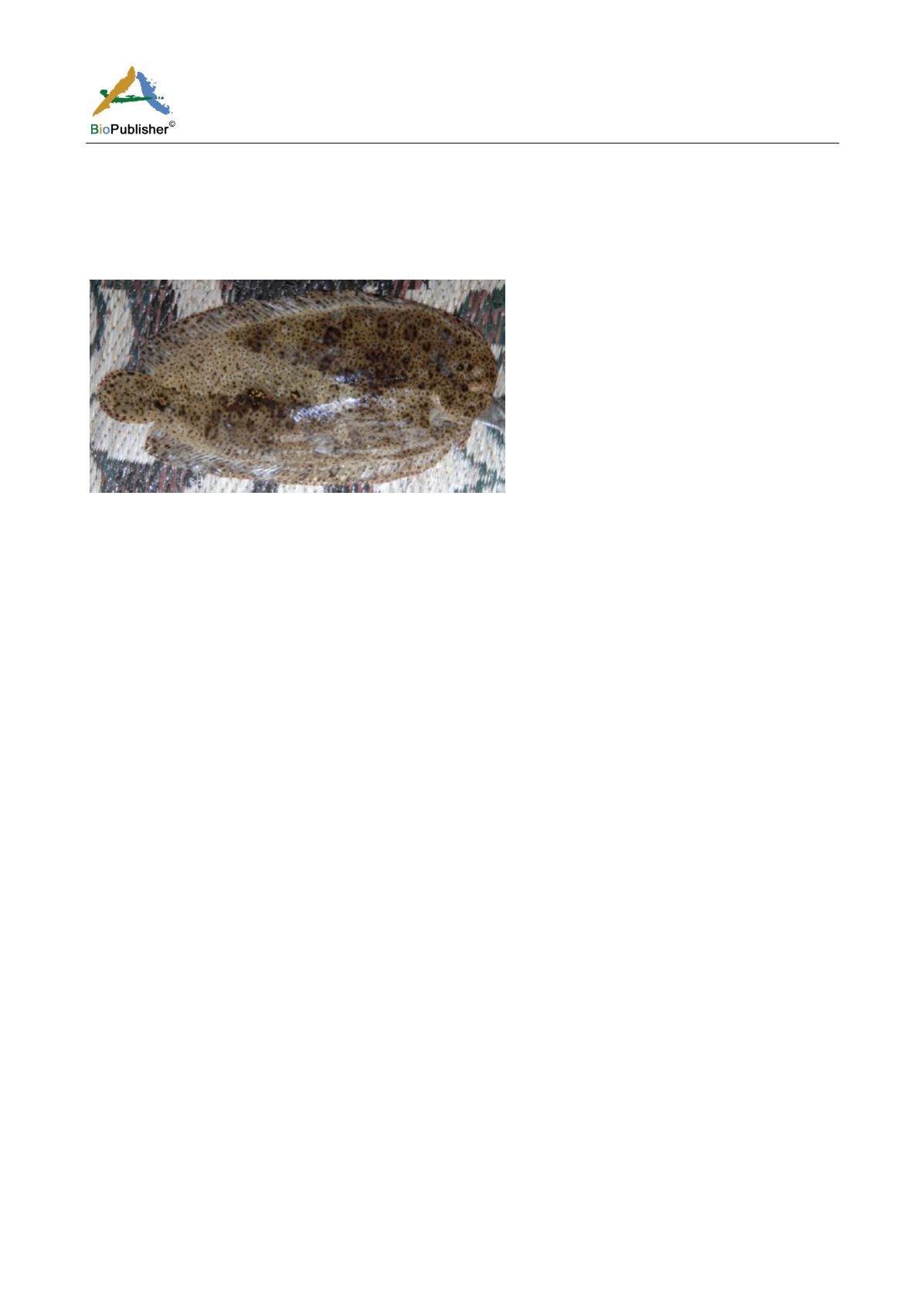
Color of abnormal specimen (370 mm TL, 359 mm SL) (Figure 2).
Figure 2
Pardachirus marmoratus
, 370 mm TL, 359 mm SL, showing ambicoloration
A broad pale band extending diagonally from the posterior part of the dorsal fin and to the area just below the
operculum, and spreading backward to cover the whole base of the anal fin. The areas with original coloration
appeared to be restricted to the head, operculum and the 2/3 of the base of the dorsal fin. Another patch of the
original coloration is found covering the posterior mid-body and over the caudal fin. Except for the small dark
spots, all other dark spots and dark edged areas that are located in the malformed location appeared to be pale.
3 Discussion
In fishes, color changes can be in two types, camouflage, which is a physiological origin and the irreversible skin
color change due to the differentiation and development of chromatophores with growth. This study is concerned
with the latter type of color change.
In the specimen of
D. pictum
, the body side shown to be the most affected area than any other parts of the fish
body. Such variation in partial melanic pigmentation is could be due to the causes stated by Roulin and Ducrest
(2011). They found that
maneuvering
of the genes of the melanocortin system or of their products will have
significant effects on a set of characters. On the other hand, Slominski et al. (2004) found that the level of activity
of the different melanocortins is correlated across tissues. Other studies indicated that with the aid of
neuroendocrine communication, the activity of the melanocortin system can be locally regulated and coordinated
(Slominski and Wortsman, 2000; Slominski, 2005; Zbytek et al., 2006) and such manipulation could vary between
tissue of the fish body (Hoglund et al., 2000).
Severe hyperplasia of dermal melanophores can accompany skin melanosis, which bring about the darkened skin.
On the other hand, hyperpigmentation in teleost fishes can cause melanophore hyperplasia. Such an effect has
been stated previously in a number of fish species (Noguera et al., 2013; Ramos et al., 2013).
It was impossible to determine the cause of the partial melanism for the specimen reported in the present study.
However, the state of the specimen studied did not support the hypothesis that melanism can be as a result of a
parasitic infestation. Hsiao (1941) reported that an Atlantic cod
Gadus morhua
probably developed melanism as a
result of having the skin heavily infested by trematode larvae. The macroscopic examination of the skin of the
specimens studied showed presence of no parasites.
The mechanism of the presence of ambicolored patches in
P. marmoratus
is different from that in
D. pictum
. The
large ambicolored patch could be resulted as abnormal differentiation of pigments cells in a limited area of
eyed-side of the fish that happened during the metamorphosis. In the normal flatfish and before the
metamorphosis, chromatoblasts are evenly distributed on the left and right epidermal sides and the eye will
gradually migrates to one side during this event. Accompanying such migration, the stem cells in the skin on the


