International Journal of Marine Science, 2017, Vol.7, No.19, 183-187
184
1 Materials and Methods
On 1
st
July 2016 one specimen of
showing pughead and saddleback syndrome deformities was
caught by a bottom trawl (44 mm mesh size in cod-end) at depth of 60 m from Gerence Bay, off Çeşme, Izmir
(38°25‟N-26°16‟E). A normal specimen was also obtained from the same fishing lot used for comparison (Figure
1A; Figure 1B). The specimens were fixed in 70% ethanol and deposited in the fish collection of the Ege
University, Fisheries Faculty (ESFM-PIS/2016-10). The skeleton of both normal and abnormal specimens were
examined by x-ray and measurements were recorded to the nearest millimetre.
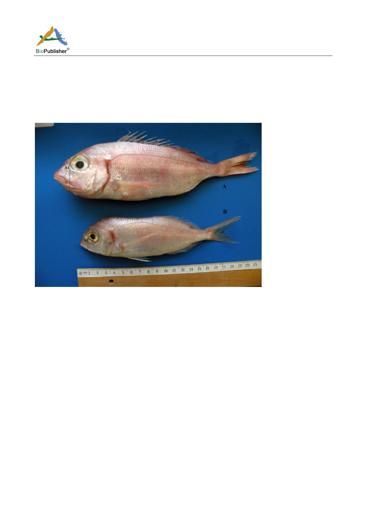
Figure
A: normal, 192 mm total length; B: abnormal, 246 mm total length
2 Results
2.1 Pughead case
The pug-headed specimen had a 192 mm total length, 153 mm standard length, 18 mm preorbital length, 33 mm
postorbital length, 15 mm eye diameter, 47 mm head length and 94 mm predorsal fin length. This specimen is
compared to normal fish having 246 mm total length, 202 mm standard length, 21 mm preorbital length, 24 mm
postorbital length and 23 and 68 mm head length.
Externally, the abnormal specimen was shown to have only short neurocranium with normal jaws and mouth was
closed, which means that the deformity has not affected the mechanism of opening and shutting the mouth (Figure
1A; Figure 1B).
The osteological deformity was compared with the normal specimen. Internally, the vomer and parasphenoid were
shortened, and displacement and/or curvature of the nasals, frontals, vomer, and palatines were observed.
Premaxilla is normal and maxilla is reduced in size. Teeth are present, but those located posteriorly on the upper
jaw appeared to be deformed in comparison with those of the normal specimen. The forehead is retracted
backward and the angle “A 2” between the line passing at the anterior edge of the eye and that passing between
the nape and the tip of the mouth is 23°, while that of the normal specimen “A1” is 46°. In comparison with the
vertebral column of the normal specimen, the contour of the 1
st
abdominal vertebra was changed in having
concave and convex upper and lower sides respectively and curved and buldging outside anterior and posterior
sides respectively (Figure 2A; Figure 2B).


