International Journal of Clinical Case Reports 2016, Vol.6, No.20, 1-7
2
1 Case Report
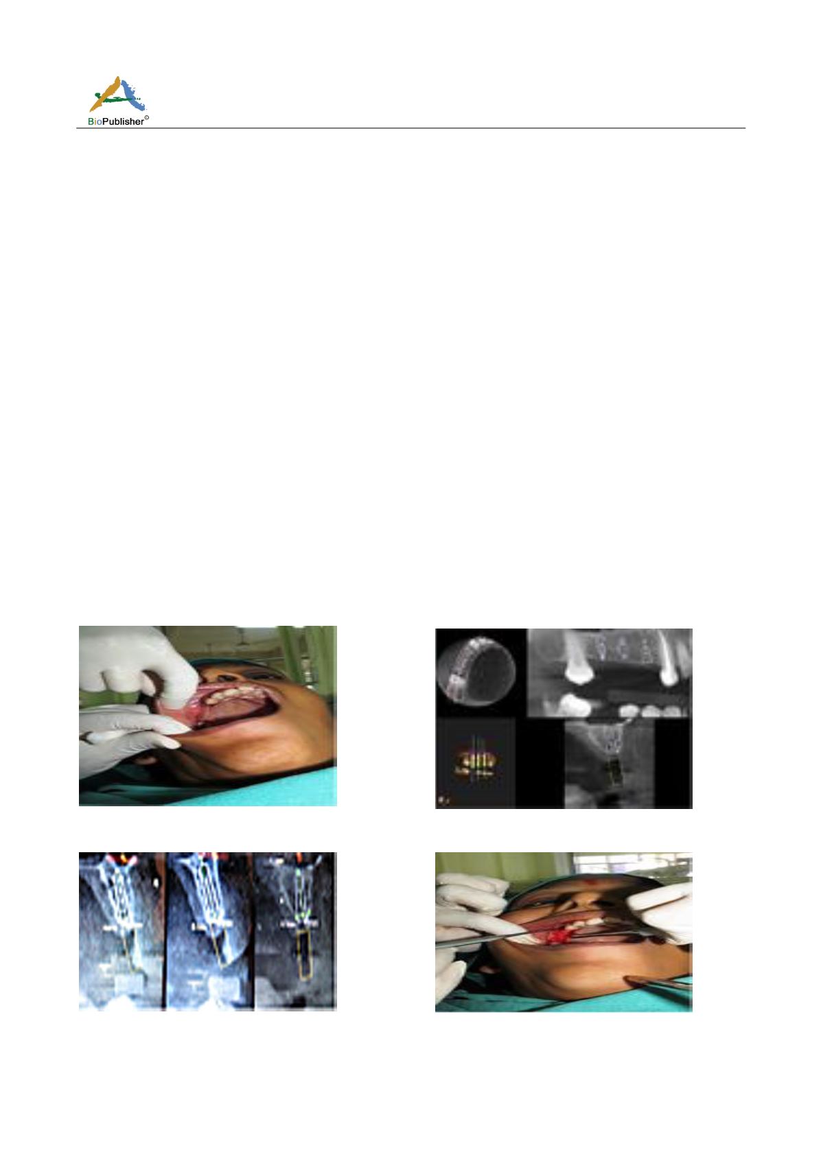
A 39 year old female patient reported to the Department of Periodontics and Oral Implantology with missing
upper right back teeth (Figure 1) since 3 years and wanted replacement of the same. All treatment options
including fixed partial denture, removable denture and implants were explained to the patient wherein the patient
opted for implant-supported rehabilitation of missing maxillary right posterior teeth. Cone Beam Computed
Tomography (CBCT) scan of 14, 15, 16 region was advised (Figure 2). Dimensions of bone available included a
mesio-distal length of edentulous segment to be of 23mm; crown height space of 5 mm in 14, 7 mm in 15 and 5
mm in 16 region; bucco-lingual width of 5.1 mm in 14, 4.6 mm in 15 and 4.7 mm in 16 region; and an alveolar
bone height of 12.3 mm in 14, 10.1mm in 15 and 8.9mm in 16 region (Figure 3). So, it was decided to place
implants of dimensions 3.75/ 11.5, 3.75/ 8, and 3.75/ 8 in the 14, 15 and 16 regions. A mid-crestal incision
followed by a crevicular incision was given in edentulous ridge in 13 till 17 region. A muco-periosteal flap was
reflected from 13 till 17 region using a periosteal elevator (Figure 4). Then, osteotomy sites were prepared using
drills in sequential manner in 14, 15, 16 region and implants were placed in respective osteotomy sites followed
by cover-screws placement (Figure 5a; Figure 5b; Figure 5c). Sutures were given to close the flap (Figure 6).
Patient was advised to take antibiotic (amoxicillin with clavulanic acid, 625 mg, TDS) and anti-inflammatory
(ketoprofen, 100 mg, TDS) for five days. Patient was also asked to rinse with chlorhexidine 012% twice daily.
Patient was re-called after a week's time for suture removal. Three months after implant placement, patient was
re-called again for second stage of the procedure and placement of healing abutments (Figure 7). The patient was
then re-called after fifteen days of the second stage procedure and impression was taken (Figure 8) and sent to
laboratory along with abutments and lab analogue. A trial was then taken after ten days and final prosthesis was
given after that (Figure 9). Splinting of implants was done for dissipation of lateral forces and better
biomechanical design. The patient was then re-called after one month of prosthetic loading to check occlusal
discrepancies, if any, and overall status of the prosthesis. IOPARs were taken at 1 month, 6 months and 1 year
follow-up visits after prosthesis delivery to check for the bony changes around implants although no bony changes
including resorptive changes were noted and patient was satisfied with the prosthesis.
Figure 1
Missing upper right back teeth
Figure 2
Cone Beam Computed Tomography (CBCT) scan
of 14, 15, 16 region
Figure 3
Dimensions of bone available
Figure 4
Muco-periosteal flap reflected from 13 till 17 region


