International Journal of Clinical Case Reports 2017, Vol.7, No.8, 33-37
34
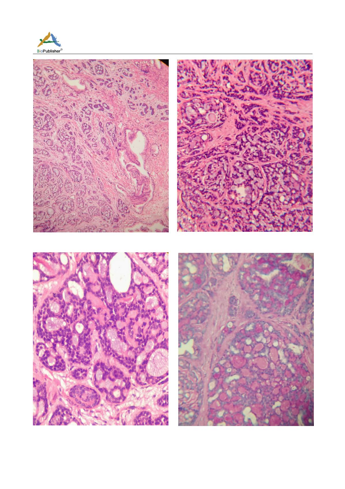
Figure 1 Scanner view showing tumor cells infiltrating normal
prostate glands. Left: tumor Right: Normal prostatic glands
Figure 2 20x view showing cribriform pattern of the tumor
cells
Figure 3 40x view showing small bland tumor cells with scant
cytoplasm and dark compact anular nuclei surround
pseudoglandular spaces with excess basement membrane and
Figure 4 20x view sowing PAS positive excess basement
membrane and mucin
mucin


