International Journal of Clinical Case Reports 2017, Vol.7, No.6, 23-27
24
habits, advanced periodontal disease, at tooth extractions sites, fungating carcinoma of buccal mucosa and patients
with tetanus with surgical opening of mouth maintained to ensure a patent airway (Sharma et al., 2008; Koteswara
and Prasad, 2010). The life cycle of the organisms starts when adult fertile flies infect the wounds and feed on
exudates and lay eggs in the injured and neurotic tissues. The first instar larvae hatch after 12-24 hrs and enter the
living tissues with feed for 5-7 days and moult twice. The third instar larvae (last stage) stop getting nourishment
from the host tissues and leave the host and pupate on the ground. Adult flies emerge after 1-2 weeks
(Maheshwari and Naidu, 2010). Incidence of oral myiasis is comparatively lesser than that of cutaneous myiasis
as oral tissues are not exposed for prolonged periods to the external environment (Kumar, 2012).
1 Case Report
A 75 year old female patient with abandoned life and of low socio-economic status presented to with a chief
complaint of swelling in the upper lip and bleeding from gums in the upper front tooth region due to worms since
4 days. Extra-oral examination revealed a diffuse swelling in the same region. On intra-oral examination, the
patient had only 18 teeth with advanced periodontal disease. Physical examination revealed larvae in the upper
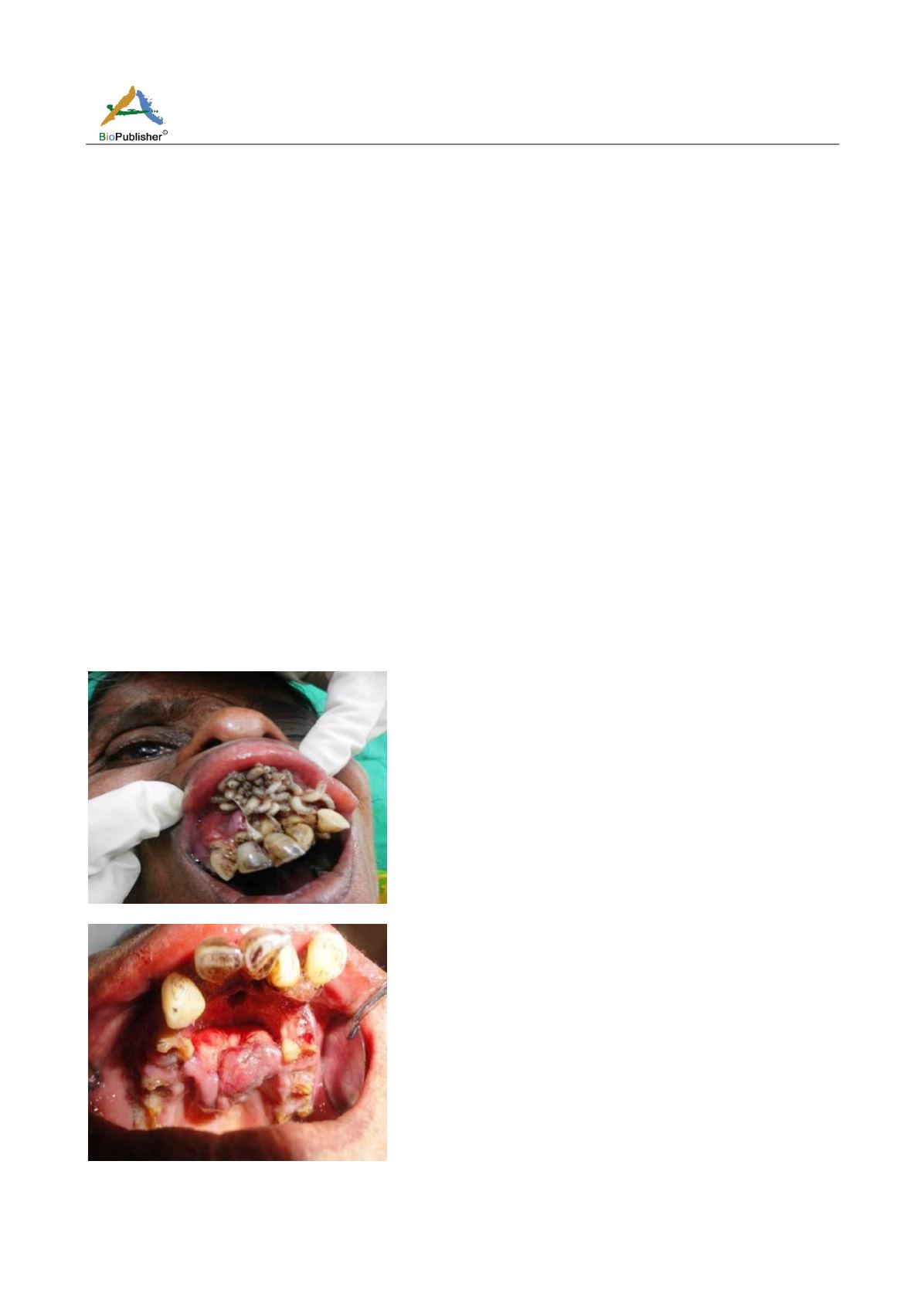
anterior teeth, both vestibular areas, subjacent to the upper lip and on the hard palate. (Figure 1) An erythematous,
ulcerative and eroded lesion was seen on upper gingiva burrowed by worms, irregular in shape. Detached attached
gingiva was present both on the labial as well as the palatal aspects. (Figure 2) Incisive canal and foramen ovale
were appreciated on clinical examination. Based on the above noted clinical findings, a provisional diagnosis of
oral myiasis was made and the patient was submitted to the removal of visible larvae. A total of more than 100
larvae were removed with the help of tissue holding forceps and the area was cleaned with betadine and saline and
the removed larvae were taken for entomological examination. Laboratory investigations were requested and an
occlusal radiograph was taken to rule-out the involvement of bone and fortunately, the infestation involved only
soft tissue. (Figure 3) HIV I and II were non-reactive and HBS antigen was found to be negative. On second visit
after three days, cotton bud impregnated with turpentine oil was applied at the site and no larvae were observed.
Figure 1 Larvae in upper anterior teeth, both vestibular areas, subjacent to the upper lip and on the hard palate
Figure 2 An erythematous, ulcerative and eroded lesion seen on upper gingiva burrowed by worms, irregular in shape; detached
attached gingiva present both on the labial as well as the palatal aspects


