International Journal of Clinical Case Reports 2017, Vol.7, No.2, 4-8
6
surface conditioning was not done. Right palatal vault was selected as the donor site. After establishing adequate
anesthesia, 2 horizontal incisions were placed at about 3 mm and 5 mm from the margin of the gingiva with the
first incision placed at 5 mm from the margin of the gingiva extending from first molar to the first premolar region
undermining a thin partial thickness flap in such a way that sufficient thickness of connective tissue graft
(1.5-2mm) could be obtained. The mesiodistal dimension of incisions was from first molar to the first premolar
region and the depth was such that sufficient thickness of palatal flap was maintained. The second incision was
carried-out parallel to the previous incision but at a distance of 3 mm from the margin of the gingiva keeping the
blade very close to the periosteum. It was extended apically up to the same depth as first incision (Figure 8).
Following this, two vertical releasing incisions were placed at the mesial and distal extensions of the horizontal
incisions to harvest the graft tissue. The graft was stored in moistened gauze (Figure 9). The donor site was
sutured with 3-0 silk suture material to control bleeding and to achieve healing by primary intention. The patient
was recalled at weekly intervals for follow-up and the healing was found to be uneventful. Suture removal was
done after two weeks. Root coverage of 4mm was achieved at the end of 3 months which improved the esthetics
of the patient (Figure 10). Patient was pleased with the outcome of re-treatment done to manage the complications
of the initial therapy.
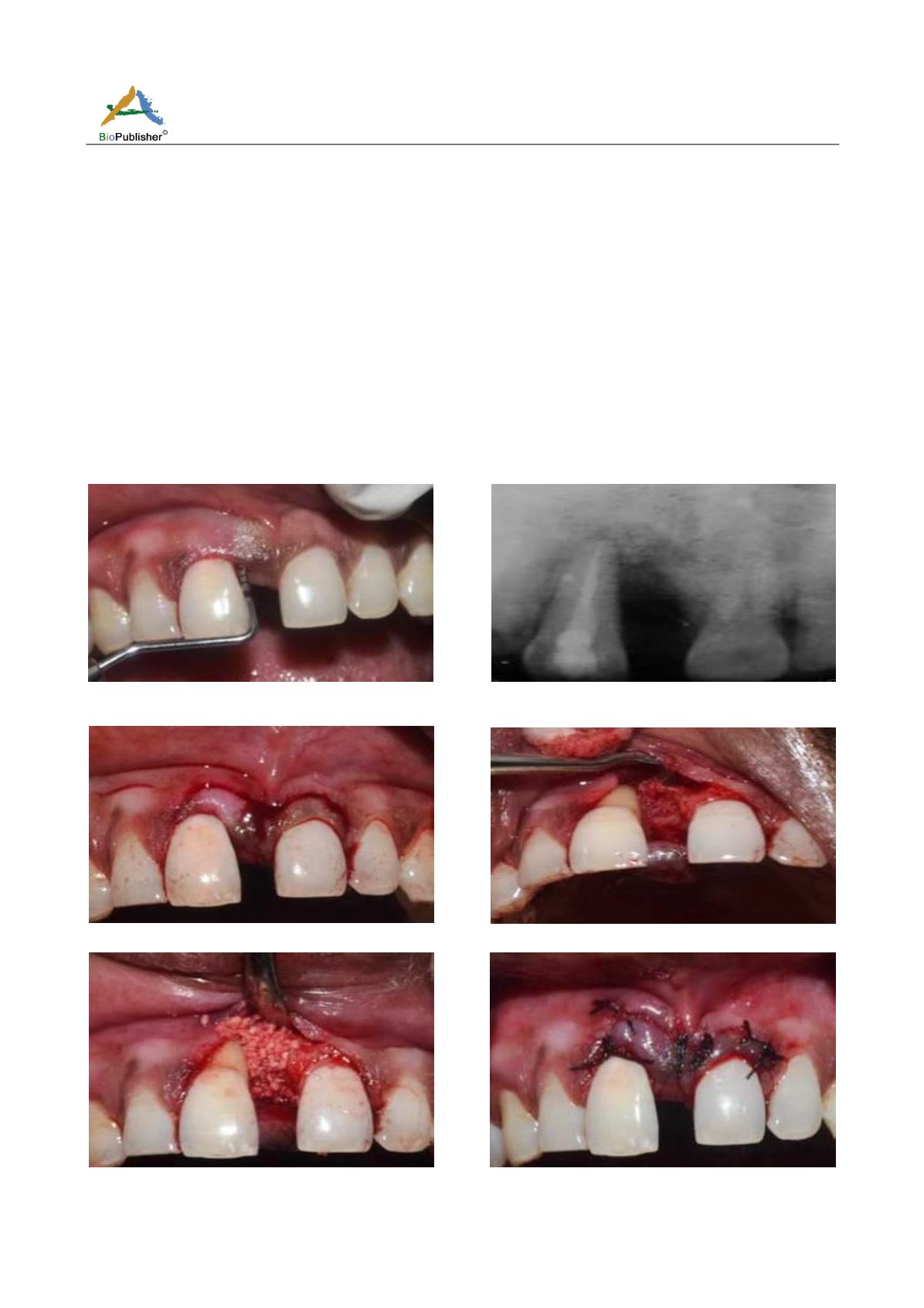
Figure 1 Pre-operative clinical photograph
F
Figure 2 Pre-operative intra-oral periapical radiograph
(IOPAR)
Figure 3 Semilunar incision
Figure 4 Exposure of the bony defect
Figure 5 Filling of the exposed bony defect with DFDBA
graft
Figure 6 Wound closed and sutures placed


