International Journal of Aquaculture, 2018, Vol.8, No.20, 151-155
154
tilapia,
O.
aureus
. These changes may lead to a disorder of lipid metabolism in the liver tissues, i.e., lipidosis,
possibly associated with toxins and extracellular products such as hemolysin, protease, elastase produced by
aeromonads (Yardimci and Aydin, 2011). In contrast, the study by Islam et al. (2008) revealed the development of
internal tissue abscess characterized by focal necrosis and haemorrhage. According to them, the distribution of
bacterial cells all over the hepatic tissue caused massive diffused necrosis represented by vacuolation and atrophy
in the liver of fish challenged with
Aeromonas.
The infiltration of haemocytes in the hepatic tissue is a measure of
cellular response, which indicated the ability of Nile tilapia to respond to the
A. caviae
infection. The results of the
present study, thus, demonstrated that
A. caviae
can cause serious pathology in the kidney, liver and pancreas of
O.
niloticus,
similar to those of other known fish bacterial pathogens such as
A. hydrophila
(Azad et al., 2001; Julinta
et al., 2017; Al-Yahya et al., 2018) or
S. agalactiae
(Adikesavalu et al., 2017).
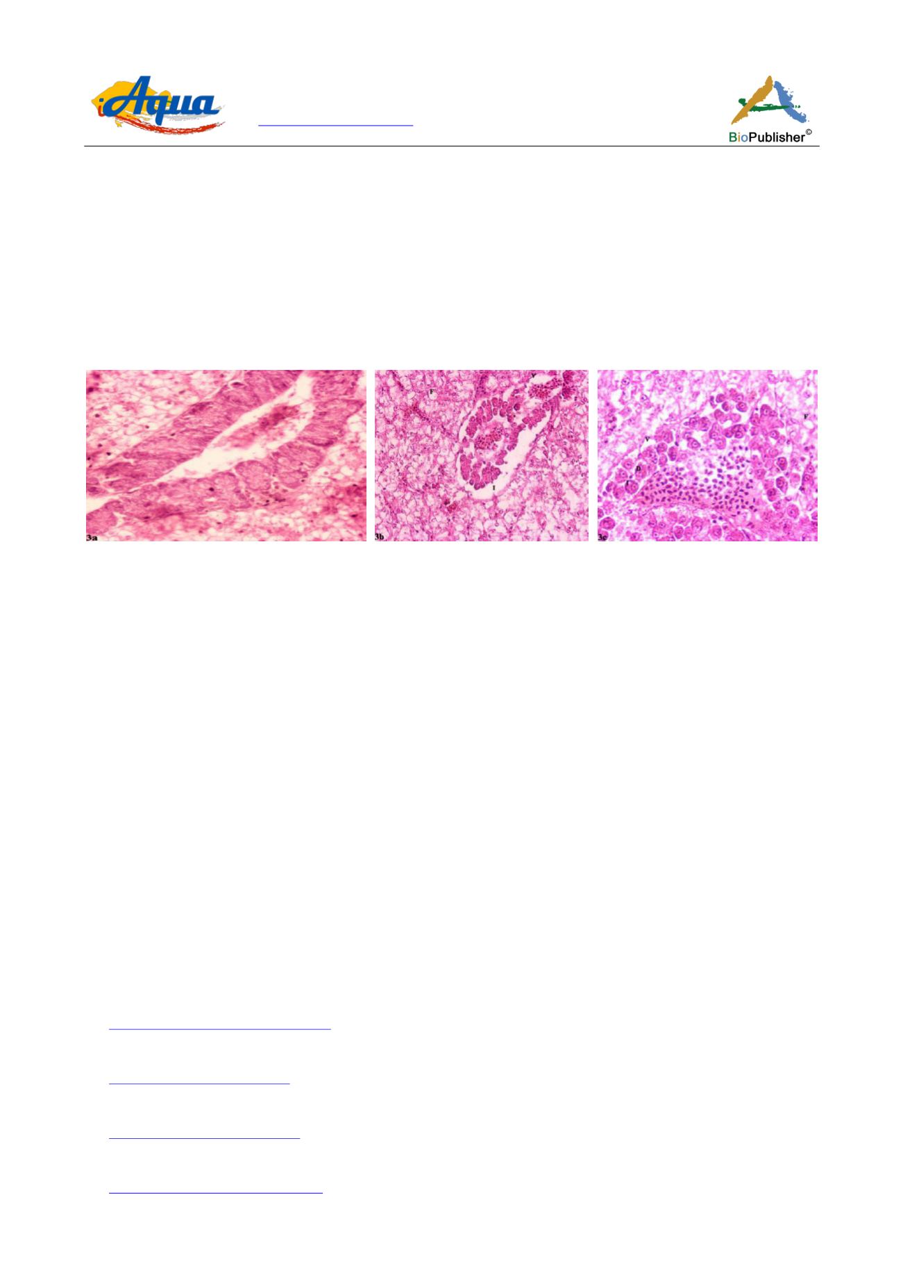
Figure 3 Photomicrography of the pancreas tissues of
Oreochromis niloticus
intramuscularly infected with
Aeromonas caviae-
T
1
K
2
showing [a] normal architecture X400 H&E staining; [b] inflamed pancreas (I), vacuolation in the pancreatic tissue (V), fatty changes
in the hepatic parenchyma (F) X400 H&E staining and [c] vacuolation (V), degradation (D), inflammation of pancreatic acinar cells
(I) and fatty changes in the hepatic parenchyma (F) X400 H&E staining
Since
A. caviae
caused mortalities only at a challenge dose of 6×10
8
cells/fish, it is highly apparent that risk
factors such as temperature fluctuations, poor water quality, accumulation of organics, crowding, etc, together
with other virulent motile aeromonads may affect the physiological functioning of tilapia and increase their
susceptibility to the pathogenic agents. Therefore, the search for timely corrective measures should first address
the identification of risk factor(s) that predispose
Aeromonas
outbreaks in Nile tilapia and mitigating the same
responsibly.
Authors’ contributions
TJA contributed to conception, analysis and interpretation of results, and write-up of the manuscript. AR and JS contributed to
sample collection, analysis, histology as well as acquisition of data. All the authors read and approved the final manuscript.
Acknowledgements
The research work was supported by the Indian Council of Agricultural Research, Government of India, New Delhi under the All
India Network Project on Fish Health (Grant F. No. CIBA/AINP-FH/2015-16 dated 02.06.2015). The authors thank the
Vice-Chancellor, West Bengal University of Animal and Fishery Sciences, Kolkata for providing necessary infrastructure facility to
carry out the work.
References
Adikesavalu H., Patra A., Banerjee S., Sarkar A., and Abraham T.J., 2015, Phenotypic and molecular characterization and pathology of
Flectobacillus roseus
causing flectobacillosis in captive held carp
Labeo rohita
(Ham.) fingerlings, Aquaculture, 439: 60-65
Adikesavalu H., Banerjee S., Patra A., and Abraham T.J., 2017, Meningoencephalitis in farmed mono-sex Nile tilapia (
Oreochromis niloticus
L.) caused by
Streptococcus agalactiae
, Archives of Polish Fisheries, 25: 187-200
Al-Yahya S.A., Ameen F., Al-Niaeem K.S., Al-Sa’adi B.A., Hadi S., and Mostafa A.A., 2018, Histopathological studies of experimental
Aeromonas hydrophila
infection in blue tilapia,
Oreochromis aureus
, Saudi Journal of Biological Sciences, 25(1): 182-185
Austin B., and Austin D.A., 2012, Bacterial fish pathogens: Disease of farmed and wild fish, (Fifth ed.), Springer-Praxis in Aquaculture in Fisheries, Praxis
Publication Ltd., Chichester, UK, pp.457


