International Journal of Aquaculture, 2017, Vol.7, No.15, 101-105
102
As far as the authors are concern, no xanthic phenotype has been on record of the silver carp from both the wild
and the artificial habitats. Therefore, the aim of this study is to document and describe for the first time ever the
presence of xanthic case for this species.
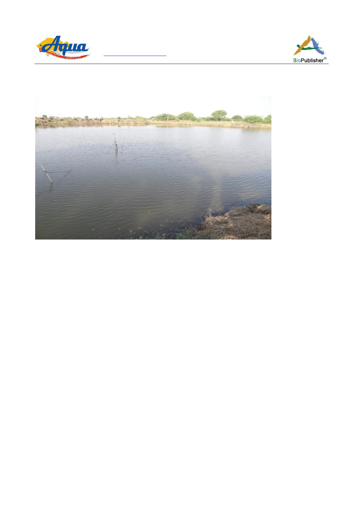
Figure 1 View of the pond of Maharashtra Fish Seed Production Centre, where the fish specimens obtained
2 Material and Methods
Two specimen of silver carp
Hypophthalmichthys molitrix
(TL 750-762 mm, SL 610-470 mm, normal specimen
TL 297 mm, SL 282 mm) were obtained on 21st November 2016 and 4th January 2017. The specimens were
supplied by Maharashtra Fish Seed Production Centre, Kesapuri camp, near Majalgaon Tahasil, Maharashtra,
India (Figure 1). It is a circular hatchery and includes one breeding pond, three spawning tank, three nursery pond,
six rearing pond and three stocking ponds. Its area is 12 acre, with pond size 150 x 100 x 6 and 180 cm maximum
depth (depth 6 feet). Source of water is from Majalgaon reservoir and wells. Body and fins were examined
carefully for external parasites, malformations, amputations and any other morphological anomalies. The
specimens were kept frozen at the Maharashtra Fish Seed Production Centre, Maharashtra, India. Once in the
laboratory, measurements were recorded to the nearest millimetre.
3 Results
Xanthic phenotype was described from two specimens of
H. molitrix
. The description of the distribution of the
xanthic pigmentations is given below based on the actual observation (Figure 2) and compared with that of the
normal specimen (Figure 3a). They have the following body proportions: total length 750, 762 mm; standard
length 610, 470 mm; head length 195, 151 mm; predorsal fin length 170 mm, 175; prepectoral fin length 200, 163
mm; preanal fin length 495, 390 mm; caudal fin length 140, 110 mm.
The whole body of the two xanthic phenotype specimens is covered with yellow color, with faint orange color on
head except the eye and all fins (Figure 3b). The dark pigmentations found on the whole body of the normal
specimen were disappeared. Instead, there were red to orange spots of different sizes were distributed in the
ventral side of the fish body. These spots have aggregated mainly in the areas of the pelvic and anal fins, and
caudal peduncle.
One of the xanthic specimen has shown a distorted lateral line. The distortion is observed in the ascending part of
the lateral line posterior to the head and in the area dorsal to the anal fin (Figure 4a; b).


