International Journal of Aquaculture, 2017, Vol.7, No.11, 79-82
80
3 Results
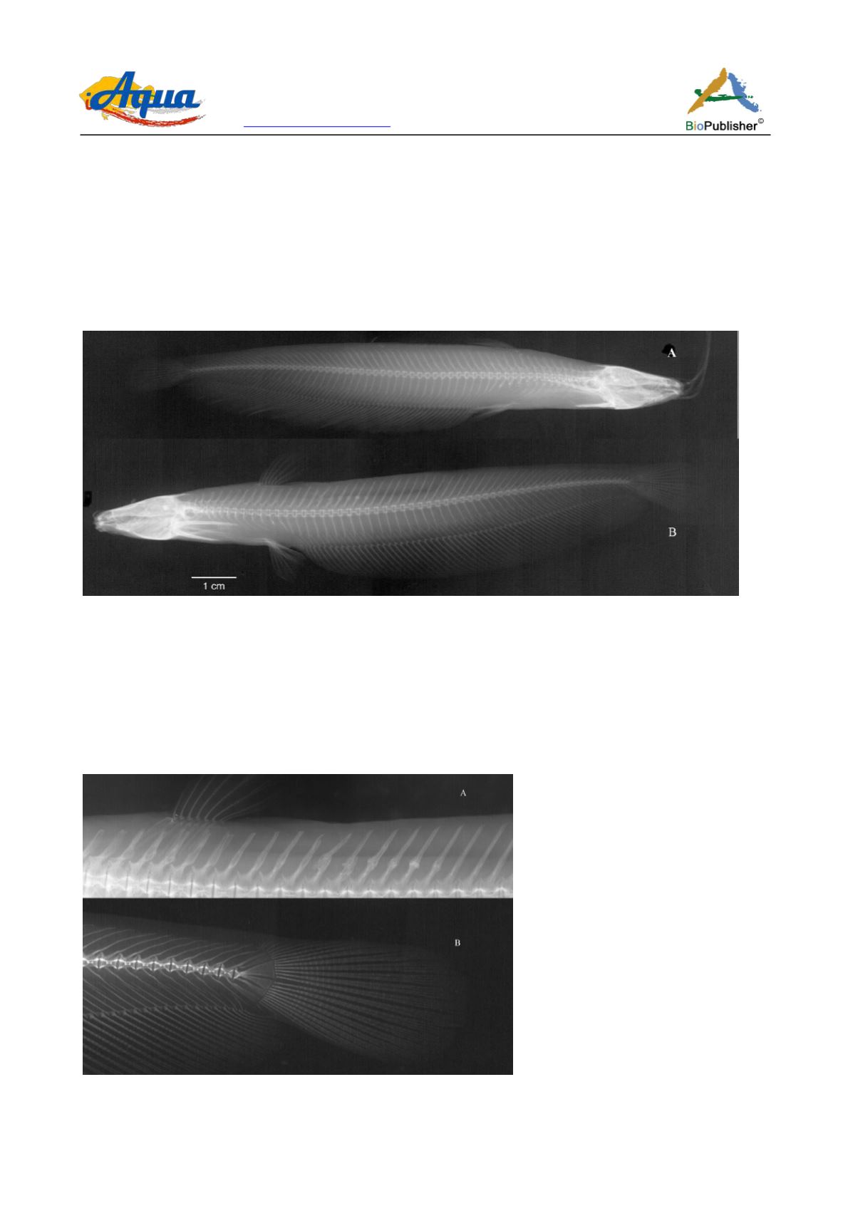
In the abnormal specimen of
H. fossilis
, there are 44 caudal vertebrae (Figure 1a; b). In those vertebrae, there are
two locations of deformities observed. The 1st location involve the neural spines of the 4
th
– 9
th
caudal vertebrae
and the 2
nd
location involves the neural and haemal spines of the 36
th
– 44
th
caudal vertebrae. The middle of the
neural spine of the 4
th
– 9
th
caudal vertebrae shown to have an abnormal ossification, where an irregular bony
lump is present. Those of the 4
th
and 5
th
vertebrae have different shape from those of the 6
th
– 9
th
vertebrae. In the
later 4 neural spines, the lump takes the spherical shape. In the neural spines of the 4
th
and 5
th
caudal vertebrae, the
ossification is irregular and covers large area, with the neural spine of the 4
th
vertebra being curved backward and
that of the 5
th
vertebra appeared as two parts joined irregularly (Figure 1a).
Figure 1 Radiograph of
Heteropneustes fossilis
showing: a, skeleton of normal specimen, 123 mm TL; b, skeleton of abnormal
specimen 137 mm TL
The 2
nd
location of the abnormality appeared in the radiograph of the abnormal specimen is related to the neural
and haemal spines of the 36
th
– 44
th
caudal vertebrae. These spines shown to be wavy instead of being straight as
in the normal case (Figure 2b). The state of waviness in the neural and haemal spines of the posterior caudal
vertebrae is more severe than those of the anterior caudal vertebrae. In the caudal vertebrae 38
th
– 44
th
, the neural
and haemal spines are wavy from their base near the centrum to the tip of the spine near the dorsal side of the fish
body.
Figure 2 Radiograph of
Heteropneustes fossilis
showing: a, close-up view of the deformed neural spines; b, close-up view of the
deformed neural and haemal spines of the caudal fin vertebrae


