Cancer Genetics and Epigenetics 2017, Vol.5, No.6, 28-32
29
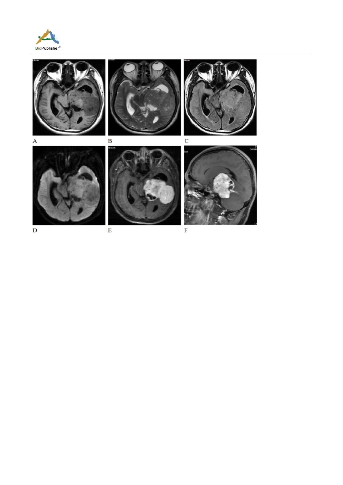
Figure 1 Preoperative MR imaging
Note: Axial noncontrast T1-weighted (A), T2-weighted (B), FLAIR (C) and DWI (D) revealed a heterogeneous expansive and
multi-lobular mass involving left ventricle. Axial and sagittal Gd-enhanced T1-weighted (E, F) demonstrated intense and
heterogeneous enhancement. Tentorium was thickened and enhanced. Small cystic changes were found in the peripheral part of the
tumor with no enhancement
1.3 Operation
We therefore performed surgical excision of the tumor. Intracranial hypertension was demonstrated during the
operation. Grossly, the mass was found hypervascular, solid and well defined, but infiltrated into the tentorium.
The cut surface was firm.
1.4 Histopathological features
The definitive pathologic examination revealed SS with uniformly spindle cells. Immunohistochemical analysis
showed VIM, p53, Ki67 (8%+) and bcl-2 positive staining. S-100, epithelial membrane antigen (EMA) and CD34
showed a negative result. The morphological and immunehistochemical features were characteristic of a
monophasic synovial sarcoma.
2 Discussion
Synovial sarcoma (SS) is generally considered a high-grade malignant neoplasm, representing between 5% and
10% of all soft tissue sarcomas (Herzog et al., 2005; Sultan et al., 2009; Shi et al., 2013). SS of the head and neck
region is quite rare, and accounts for approximately 3% to 10% of all synovial sarcomas. The most common site
for SS in the head and neck region is the hypopharynx, because it is the seat of numerous synovial formations. To
the best of our knowledge, this is the first reported case of SS of the intraventricle involving tentorium.
The origin of SS is still controversial. Virtually, it is named exclusively for its appearance, and as SS neither arise
from nor differentiate toward synovium, the name is a historical error (Smith et al., 1995; Fisher et al., 1998;
Thwayet al., 2014). Owning to its more common origination from the pluripotent mesenchymal cells nearby or
even remotely from articular surfaces, tendons, tendon sheaths, juxta-articular membranes, and facial aponeuroses,
rather than from mature synovial tissue. The ventricular SS, which has not been reported yet, is quite a unique site
for the occurrence.


