International Journal of Clinical Case Reports 2015, Vol.5, No. 39, 1-3
2
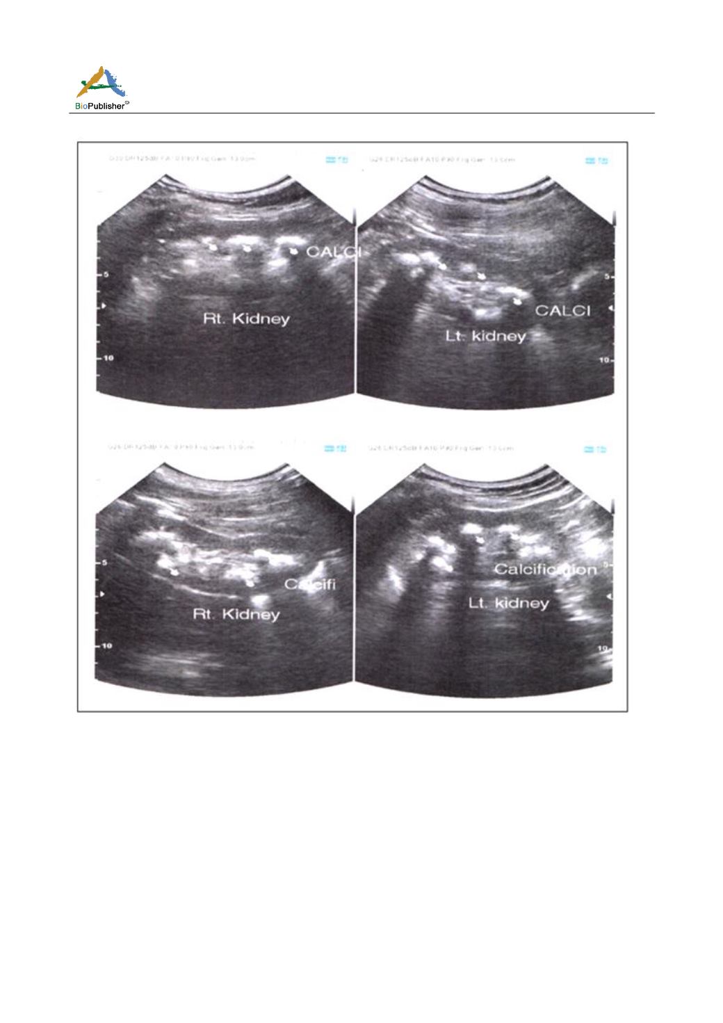
Figure 1 Ultrsonogram of whole abdomen
revealed normal calcium and phosphate level along
with elevated PTH level (611.8 ng/ml). Thereafter we
focused on her neck glands. Ultrasonogram(USG) of
neck (Figure 2a) showed solitary hypervascular nodule
near lower pole of right thyroid (1.8x1.4 cm) with CT
scanning (Figure 2b) later on showing the nodule to be
a parathyroid adenoma (2x1 cm). It was nonmetastatic
although some prominent cervical lymph nodes were
there, mostly reactive. Parathyroid scintigraphy (sestamibi
scan) concluded it was parathyroid adenoma or
hyperplasia and simultaneously they advised to rule
out hot nodule of thyroid. So we ran thyroid function
test which was quite normal. Her urine calcium &
urine calcium-creatinine ratio was found to be elevated
which excluded familial hypocalciuric hypercalcaemia.
To find out any skeletal changes owing to hyperpara-
thyroidism we performed x-ray of both hands and skull
which were normal. But Dual Energy X-ray
Absorptiometry (DEXA) showed T score of AP spine,
femur and radius below -2.5. Her endoscopy of upper
GIT was also normal.


