Basic HTML Version


International Journal of Molecular Veterinary Research
2013, Vol.3, No.4, 9-12
http://ijmvr.sophiapublisher.com
10
much easier to be seen on ultrasonogram as compared
to radiograph. Therefore the shape of calculus on
ultrasonogram will depend on the density of
surrounding tissue i.e. fluid or soft tissue. The change
in urine density can also be seen by ultrasonography.
The treatment for presence of calculus in urinary
system is carried out by both methods i.e. surgical and
medicinal. The urethroliths and cystoliths are best
managed by surgical methods (Kyles et al., 2005). In
present study also it was observed that after surgical
removal of calculus from urethra and urinary bladder
the recovery of dogs was uneventful. The nephroliths
can be managed by medical management also (Caraza,
2005) i.e. by using lithotripsy (Davidson et al., 2003),
and dietary management (van Metre et al., 1996). In
this study also the nephroliths were successfully
managed by providing oral urinary antiseptics such as
cystone. From this study it can be concluded that the
use of fluid, antibiotics, analgesics, anti-inflammatory
drugs, B-complex, oral urinary antiseptics i.e. cystone
tablets after surgery were found to be useful for
control of recurrence of lodgment of any calculus in
urinary system. In dogs radiography is observed to be
better diagnostic technique than sonography in cases
of cystoliths and urethroliths. The sonography is
comparatively better technique than radiography for
nephroliths. The change of urine density in urinary
bladder can be better observed by sonography.
2 Materials and Methods
Ten dogs, seven male and three female between age
group of 5~7 years were admitted to the teaching
veterinary hospital with the history of difficulty in
urination. In these dogs, initially there was dribbling
of blood tinged urination from the urethral orifice but
in later stages either the urination was drop wise or
there was complete stoppage of the urination. Several
drugs such as diuretics, antibiotics, anti-inflammatory,
B-complex and fluids were tried by the local
veterinarians but there was no response. There was
frequent vomition and constipation also. These dogs
were dull, depressed and dehydrated. Polyethylene
catheter was tried through urethra to take the urine out
but it could not be passed. Immediately these dogs
were referred for radiographic and ultrasonographic
examinations. Blood was also collected and sent for
laboratory examination. Hematology revealed mild
anemia in these cases. There was increase in TLC,
DLC and Packed Cell Values. Biochemical evaluation
showed increase in values of Blood Urea Nitrogen,
Serum Creatinine, and SGOT and SGPT.
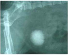
Radiographic reports indicated multiple cystoliths and
urethroliths. In three male and two female dogs there
was a single large sized calculus present in the center
of the urinary bladder (Figure 1) while there was no
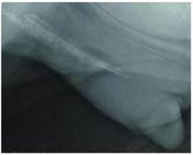
calculus present in the urethra. In three other cases,
there was unusual big calculus in the centre and
many small calculi at the bottom of the urinary
bladder. In these dogs almost entire urethral passage
was occupied by the small calculi present in
multiple rows (Figure 2).
Figure 1 Big cystolith
Figure 2 Multiple urethropliths
Ultrasonographic picture of the kidney, urinary
bladder and urethra showed presence of cystoliths,
urethroliths and nephroliths (Figure 3). The
nephroliths could not be seen by radiography. A
hyperechoic half moon shaped image of the cystolith
was observed in the urinary bladder surrounded by the
hypoechoic area. Density of the urine was appeared
cloudy. The shape and size of cystoliths were observed
to be changed due to acoustic interface (Figure 4). The
urethroliths could not be seen clearly on