Basic HTML Version


International Journal of Marine Science 2014, Vol.4, No.44, 1-14
http://ijms.biopublisher.ca
3
to environmental pollution in each of the 3 species.
We measured enzyme activities (SOD, CAT, PER, GR,
ALT, AST) and concentrations of oligopeptides,
albumin and hemoglobin in examined elasmobranch
tissues. These data provide baseline information
against which comparisons can be made in monitoring
the adaptability and response of elasmobranchs in a
changing marine ecosystem.
2 Materials and Methods
2.1 Capture and sampling
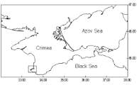
Biological sampling was performed in the coastal
waters of Sevastopol (Figure 1). Three elasmobranch
species Atlantic spiny dogfish (
Squalus acanthias
,
n=16), buckler skate (
Raja clavata
, n=43) and
stringray (
Dasyatis pastinaca
, n=31) were studied in
summer period of 2010-2013. Animals were captured
by the fishery and immediately transported to the
laboratory in the containers with marine water and
constant aeration. After blood sampling the animals
were decapitated.
Figure 1 Sampling sites of 3 species of elasmobranchs in
Sevastopol coastal waters (44°36’N-33°32’E, Sevastopol,
Black Sea, Ukraine)
Fish were individually measured and weighed. The
total length (from tip of nose to tip of tail), standard
length (from tip of nose to precaudal pit.) and total
weight were measured according the methods
described Pravdin (1966).
2.2 Blood collection, processing, and analysis
Blood (approximately 1~3 mL) was collected from the
ventral tail artery using needle syringe or Pasteur
pipette. Whole blood was collected and serum was
separated within 24 hrs of collection in refrigerator at
4
℃
.
After blood collection fish were dissected and the liver
was quickly removed and stored on ice. The organ
was washed in the cold 0.85% NaCl solution several
times, then homogenized in a physiological solution
(1:5 w/v) using glass homogenizer. The resulting
homogenate was centrifuged at 8000 g for 20 min.
Biochemical assays were performed immediately after
liver preparation.
Specifically, the sediments of red blood cells (RBC)
were washed three times with cold 0.85% NaCl
solution and then lysed by addition of 5 vol of
distilled water and stored for 24 hrs at 4
℃
as we
described previously (Rudneva, 1997). The enzyme
activity was then determined in the RBC lysates
immediately after preparation. Spectrophotometer
Specol-211 (Carle Zeiss, Germany) was used for all
biochemical determinations.
2.3 Biochemical assays
Oligopeptide concentrations (OP): The concentration
of oligopeptides (OP) was detected separately from
RBCs, serum and liver extracts of the 3 species.
Specifically, 0.25 mL Trichloracetic acid (TCA) was
added to 0.5 mL of the sample and centrifuged at 9000
g for 30 min. Next, 0.3 mL of the supernatant was
mixed in 3.7 mL 3% NaOH and 0.2 mL Benedict
reagent. The mixture was incubated for 15 min at
room temperature and then optical density (OD) was
measured at 330 nm (Karyakina and Belova, 2004).
We express the results in arbitrary units (OD at 330
nm per mg protein ×10
-3
).
Antioxidant activity (SOD, CAT, PER, GR):
Antioxidant activities in the liver extracts and red
blood cells from the 3 species in this study were
determined according to methods described previously
(Rudneva, 1997), with a few minor modifications.
Specifically, Superoxide dismutase (SOD) was
assayed on the basis of inhibition of the reduction of
nitroblue tetrasolium (NBT) with NADH mediated by
phenazine methosulfate (PMS) under basic conditions
(Nishikimi et al., 1972). All measurements were
performed in 0.017 M sodium pyrophosphate buffer
pH 8.3 at 25
℃
. The reaction mixtures contained 5 µM
NBT, 78 µM NADH, 3.1 µM PMS, and a 0.1 mL
Black
Sea
Sevastopol
skaya Bay
▄ -
sampling sites