Basic HTML Version


International Journal of Marine Science 2013, Vol.3, No.38, 306-310
http://ijms.sophiapublisher.com
308
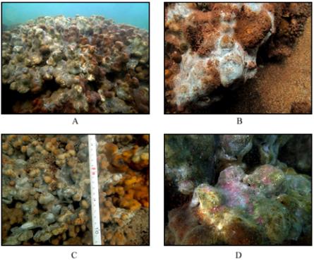
Figure 3 A: Mass mortality of
Porites
colonies at Hormuz
Island due to bacterial mats B: White mat on a
Porites
colony C:
White mats changed overlying black layers due to iron sulfide
precipitation D: Photosynthetic sulfur and non-sulfur bacteria
are probably responsible for pink and green colors
Reports on post-bleaching coral mortality due to
diseases are increasing worldwide (Bruno et al., 2007;
Miller et al., 2009; Riegl et al., 2011; Bastidas et al.,
2012). Although, coral reefs facing mild and
sometimes severe bleaching can recover quickly (e.g.
Goreau et al., 2000; West and Salm, 2003; Riegl et al.,
2011), diseases can reduce resilience, coral cover,
and reef resistance drastically for several years
(Goreau et al., 2000; Rosenberg and Loya, 2004;
Sutherland et al., 2004).
Massive
Porites
corals are known as the most tolerant
corals to thermal stress (Goreau et al., 2000; Loya et
al., 2001); however, the results of this study indicate
Porites
corals are still susceptible to the secondary
effects of bleaching events including coral diseases.
Moreover, reefs affected by coral diseases have less
resistance and resilience (Goreau et al., 2000;
Rosenberg and Loya, 2004) resulting in more
likelihood of being overgrown by invasive organisms
and competitors such as macroalgae and other reef
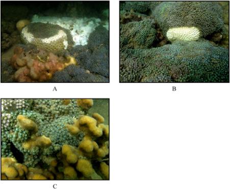
builders; however, even under no visible stress,
zoanthids are able to overgrow reef-building corals
(Figure 4A, B, C; J. Kavousi, personal observation).
The reefs to the east of Hormuz Island are now
dominated by zoanthids (59.79±15.95%). Whereas
reef-building corals were highly affected by the recent
bleaching event and its consequences, zoanthids
showed no sign of bleaching or sickness. The shift
Figure 4 Overgrowth of zoanthids on coral colonies including
A:
Favia
B:
Platygyra
C:
Porites
at the east of Hormuz Island
from coral dominated reefs to non-scleractinian
coral-dominated reefs due to climate change and its
consequences were reported (reviewed by Norström et
al., 2009; Bell et al., 2013). This may lead to local
extinction of reef-building corals of the east of
Hormuz Island under ongoing climate change.
Sulfide oxidizing bacteria such as
Beggiatoa
,
Thiothrix
and
Thioploca
, etc. are suggested to be the
dominant bacteria in the white mats (Jorgensen, 1977;
Jorgensen and Postgate, 1982; Fenchel et al., 2012). A
dark colored underlayer (Figure 3C) that appeared at
the white surface of affected tissues less than 24 hours
after the first observations is probably due to iron
sulfide precipitation. The pink and green colored
underlayers observed on the majority of infected coral
colonies (Figure 3D) could be photosynthetic sulfur
and non-sulfur bacteria; however, microbial examinations
are needed.
Although sulfide oxidizing bacteria linked coral
mortality was reported (Garrett and Ducklow, 1975;
Mitchell and Chet, 1975), previous observations
involved very localized coral mortality, often due to
artificially induced stress in the laboratory or linked to
sediment stress in the field (Weber et al., 2012).
Sulfide oxidizing bacteria are a visible epiphenomenon
that is a result, not a cause, of mortality. Coral surface
tissue smothered with fine-grained mud creates locally
anoxic sites (Erftemeijer et al., 2012) that are
colonized by anaerobic, heterotrophic sulfate-reducing
bacteria. The Hydrogen Sulfide they produce then