International Journal of Marine Science, 2017, Vol.7, No.32, 308-315
312
mouth, a broad patch located below the spinous part of the dorsal fin and beneath the pectoral fin. It extended
ventrally to reach the dorsal edge of the ventral fin, a nearly rectangular patch lie at the base of the junction of the
spinous and the soft parts of the dorsal fin. Ventrally it extended to the lateral line, a nearly rectangular patch
situated below the soft part of the dorsal fin and extended posteriorly to reach the posterior edge of the dorsal fin.
It extended ventrally to reach the lateral line and small rectangular patch located dorsal to the base of the anal fin
and traverse posterior ventrally on the surface of the anal fin. Few other small patches of the normal coloration
were observed on the dorsal side of the head, below the anterior edge of the dorsal fin, below the operculum and
at the base of the caudal fin. The caudal fin showed to be color aberrated (Figure 6). The right side of the fish was
less aberrated than the left side. Here, big patch in the shape of backward reversed “L-shaped” was located from
the nape to the anterior edge of the dorsa fin. It extended ventrally to reach the area from the operculum to the
anterior end of the anal fin. Pale narrow patches were found along the posterior ventral edge of preoperculum and
operculum. Few small patches of the normal coloration were found on the caudal peduncle. The caudal fin showed
to be color aberrated (Figure 7). The ventral side of the fish body showed small dark patches on the gular area and
under the pectoral fins (Figure 8).
Plectorhinchus playfairi
Colour of normal specimen (Figure 9):
The color of the body of this species is distinguished in having the dorsal side black and white ventral side.
Presence of 4 white vertical bars, 1 across the operculum and 3 extending below the spinous part of the dorsal fin.
All fins were black.
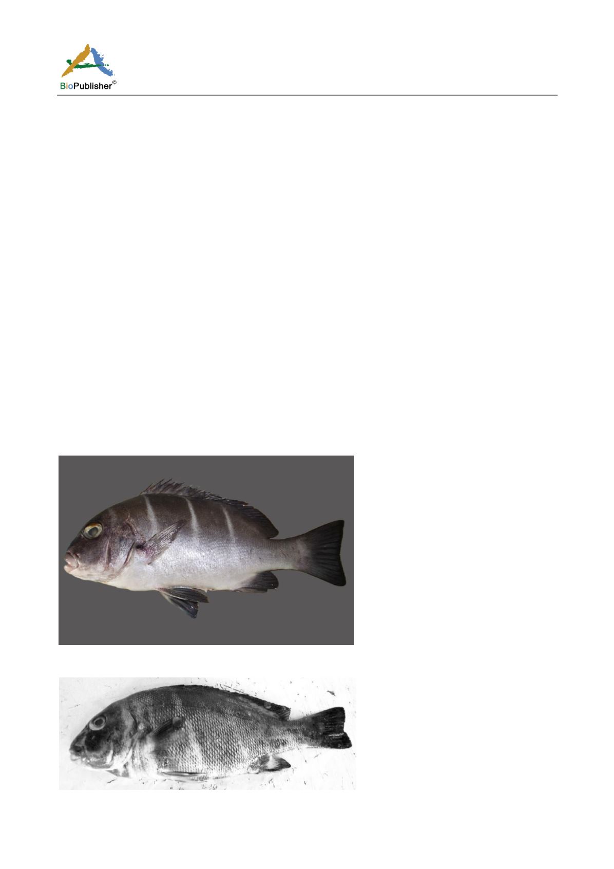
Colour of abnormal specimen (TL 346 mm) (Figure 10):
The colour of this specimen was not heavily aberrated. The areas under the operculum, the base of the pectoral fin
and the base of the caudal peduncle were shown to have pale patches. The 1
st
white bar appeared disturbed with a
pale patch.
Figure 9
Plectorhinchus playfairi
, 348 mm TL, showing normal body coloration
Note: Courtesy of Marine science and Fisheries Centre, Ministry of Agriculture, Oman
Figure 10
Plectorhinchus playfairi
, 346 mm TL, left side


