International Journal of Clinical Case Reports, 2016, Vol.6, No.26, 1-4
2
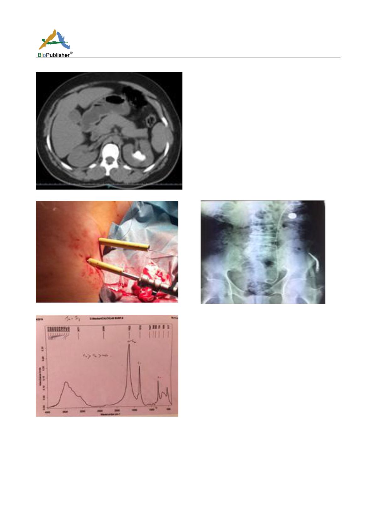
Fig.1. CT urography: left coralliform lithiasis (40mm) in the pyelic region
Fig.2. Percutaneous nephrolithotomy in two stages
Fig.3.The morphoconstitutional results of the calculi: calcium oxalate structure
As part of the aetiological investigation, hypercalcemia to 119 mg/l (88 -101), with hypophosphatemia to 14mg/l
(27 -45), PTH=508.7pg/ml (15 -65), so the diagnosis of the PHPT was established. The ultrasound cervical scan
showed a right heterogeneous hypoechoic parathyroid mass measuring 46x34 mm in contact with the thyroid
gland. The parathyroid sestamibi scintigraphy confirmed an isolated enlarged lower right parathyroid gland
measuring 5cm. A cervical MRI did not reveal any extension to the surrounding vascular structures. However,
there was an extension to the upper mediastinum in relation to the carotid artery. A conventional cervicotomy was
performed for the removal of the parathyroid adenoma which measured 5 x 2.5cm with an ipsilateral thyroid


