International Journal of Clinical Case Reports 2017, Vol.7, No.5, 19-22
20
upper left posterior regions. The gingiva seemed to be enlarged with Grade II enlargement. The gingival
enlargement was diffuse, sloppy in appearance, firm and fibrotic, accompanied by inflammation (Figure 1a).
Orthopantomograph (OPG) revealed horizontal bone loss in the said regions of maxilla and mandible (Figure 1b).
There was no drug or, systemic history reported. Differential diagnoses included diabetes and other systemic
diseases associated periodontitis which were ruled-out after determination of completed blood counts and HbA1c
levels. The treatment plan was explained to the patient and a written consent was obtained. On day 1, scaling and
root planing was performed and oral hygiene instructions were given to the patient (Figure 2). After 4 weeks of
phase I therapy, based on the amount of tissue remaining and probing pocket depth, the treatment planning was
done. For upper anterior and right posterior regions, Kirkland flap was performed for debridement whereas for
lower anterior region, modified Widman flap procedure was performed to excise the tissue along with the pocket
lining. Following this the residual plaque and calculus was removed and thorough root planning was done (Figure
3a; Figure 3b; Figure 3c; Figure 3d; Figure 3e; Figure 4a; Figure 4b; Figure 4c; Figure 4d). Periodontal Coe-pack
dressing was given and the excised tissue was sent for histopathological examination. H & E staining showed
hyperplasia of stratified squamous epithelium and an intense fibroblastic activity in the connective tissue along
with chronic inflammatory cell infiltration, chiefly composed of plasma cells and lymphocytes with numerous
blood vessels (Figure 5) suggestive of inflammatory fibro-epithelial hyperplasia. Post-operatively, patient was
given antibiotics, analgesics and chlorhexidine mouth wash. Suture removal was done after 1 week and the patient
was instructed to maintain oral hygiene. Patient showed uneventful healing after 1 month and was followed-up for
next 6 months with successful outcome.
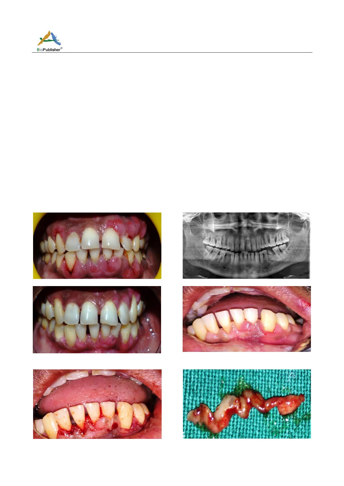
Figure 1a Pre-operative clinical photograph
Figure 1b Pre-operative orthopantomograph (OPG)
Figure 2 Clinical photograph after scaling and root planing
(subgingival curettage)
Figure 3a Clinical photograph at baseline
Figure 3b Clinical photograph after modified Widman's
incision
Figure 3c Excised tissue specimen


