Basic HTML Version


Molecular Plant Breeding 2011, Vol.2 No.2
http://mpb.sophiapublisher.com
9
the short period.
1 Results and analysis
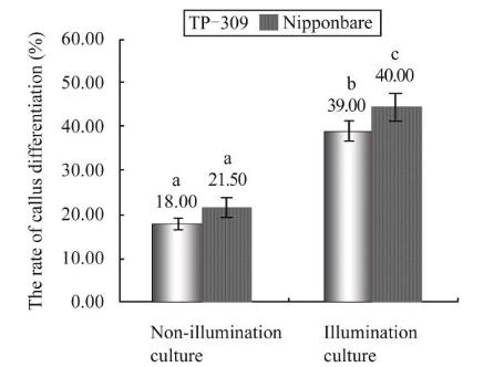
1.1 Effect of different light culture conditions on
the rate of callus differentiation
There were significantly different callus differentiation
rates existed in the two tested varieties under light
culture and non-light culture (<0.01) (Figure 1), which
indicated that light culture has promotive effects on
callus growth. We observed that callus of two varieties
after three weeks non-light culture were getting dark,
few granules appearing, callus surface drying and low
rate of callus differentiation (18% and 21%, respectively),
whereas under light culture 3 weeks, callus showed
health growth in size and color.
Figure 1 Effect of different light culture coditions on the rate of
callus differentiation
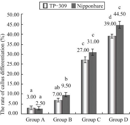
1.2 Effect of drying process on the rate of callus
differentiation
It was reported that japonica rice callus with properly
dehydrating treatment could promote rice regeneration
(Masayoshi, 1992). In this research, Effects of
dehydration treatment following infection and co-culture
on rates of callus differentiation were analysed with
the fixed dry time. The results were shown in Figure 2.
It was significant that the rates of callus differentiation
between Taipei 309 and Nipponbare were difference
among four treatment groups based on multiple
comparesions, A higher rates of callus differentiation
came out for dehydration treatments both following
post-infection and co-culture (Figure 3).
Figure 2 Effect of dehydration treatment on the rate of callus
differentiation
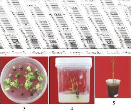
Figure 3 Selection of phosphinothricin-resistant callus and
plant regeneration
Note: 1: Callus induced from rice mature embryo; 2: The callus
cultured after 21 days light culture; 3: The callus screened by
PPT resistance; 4: The rooting plant with PPT-resistance; 5:
regeneration of PPT-resistant plants
1.3 Transgenic plants detected by histochemical
staining and PCR test
GUS histochemical staining of the resistant rice plants
were carried out in this research (Figure 4). It is easy to
see the blue spots in transgenic leaf and root, whereas
nothing in the control. Meanwhile we selected one
transformed rice plant from 370 resistant plants for pcr
test, a 875 bp length band were amplified from the