Basic HTML Version


Molecular Pathogens
MP2011, Vol.2, No.2
http://mp.sophiapublisher.com
- 11 -
size) showing a tail of 230, which might be
responsible for the disease, were described as possible
agents of the disease (Serrano et al., 2004). Double
stranded RNAs (dsRNA) with a size ranging from 0.6
to 6.6 kb were obtained from infected trees in Turkey
(Açýkgöz and Timur Döken, 2003). The main
objective of the present work is to identify and
characterize an Egyptian isolate of the
fig mosaic virus
(FMV).
1 Results
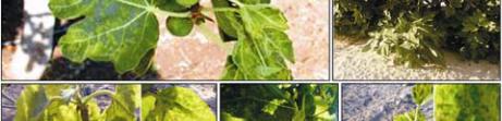
1.1 Disease Incidence
Fig mosaic symptoms were observed in all the fields
surveyed. The symptomatology identification of FMV
was recorded as: light chlorotic spotting, mottling,
extensive chlorosis along the veins and leaf
malformation as shown in Figure 1.
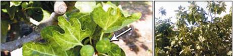
Figure 1 The observation of Fig mosaic symptoms
Note: Overview of fig (
Ficus carica
L.) plants showing
virus-like symptoms at different locations; Trees showing FM
symptoms i.e. on leaves different patterns of chlorotic to
yellowish mottling and various types of leaf deformation, on
fruits chlorotic spots were very similar to those on leaves
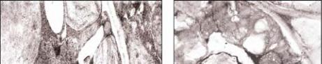
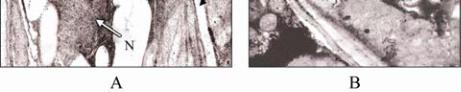
1.2 Electron microscopy and Cytopathological
effects
Electron micrographs of ultrathin sections prepared from
healthy
F. carica
L. leaves, represented in Figures 2,
exhibited normal cell structures, while those prepared
from FMV-infected
F. carica
L. leaves revealed many
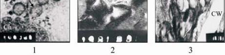
cytopathological effects. Figures 3 show starch grains
accumulation inside the chloroplasts. Two types of
intracytoplasmatic electron-dense bodies with a double
membrane (DMBs) were observed in parenchyma and
subepidermical cells of fig mosaic leaves, always
presented in rounded to ovoid 160~200 nm in size and
elongated, straight to slightly flexuous up to or exceeding
1µm in length. Long elongated and flexuous virus-like
particles surrounding the chloroplast in parenchyma cell
are shown Figure 4.
1.3 Detection of
fig mosaic virus
using the
NIB
gene
universal primer of
Potyviruses
Universal primers of the Nuclear Inclusion Body of
the
potyviridae
were designed by (Chen et al., 2001)
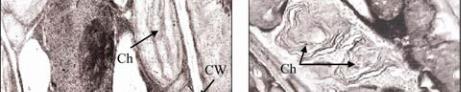
Figure 2 The results of electron micrographs of ultrathin
sections from
F. carica
L.
Note: A: Electron micrographs of ultrathin sections from
healthy leaf of
F. carica
L. showing normal cell structure; B:
Infected leaf of
F. carica
L. showing deformations of
chloroplatides; (CW): cell wall; (N): nucleus; (Ch): Chloroplast;
X= 22 000
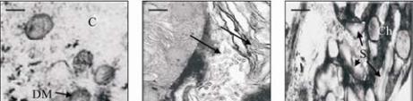
Figure 3 Starch grain accumulation by electron micrographs of
ultrathin sections
F. carica
L.
Note: A: Electron micrographs of ultrathin sections from
FMV-infected leaf of
F. carica
L. showing starch grain
accumulation; B: Elongated, slightly flexuous DMBs in
parenchyma cell. C: A group of globose double membrane
bodies (DMBs) in parenchyma cell (C) cytoplasm; (Ch)
Chloroplast and (S) starch grain, (CW) cell wall; X in A=25
000; B and Care 50 000